Figure 3.
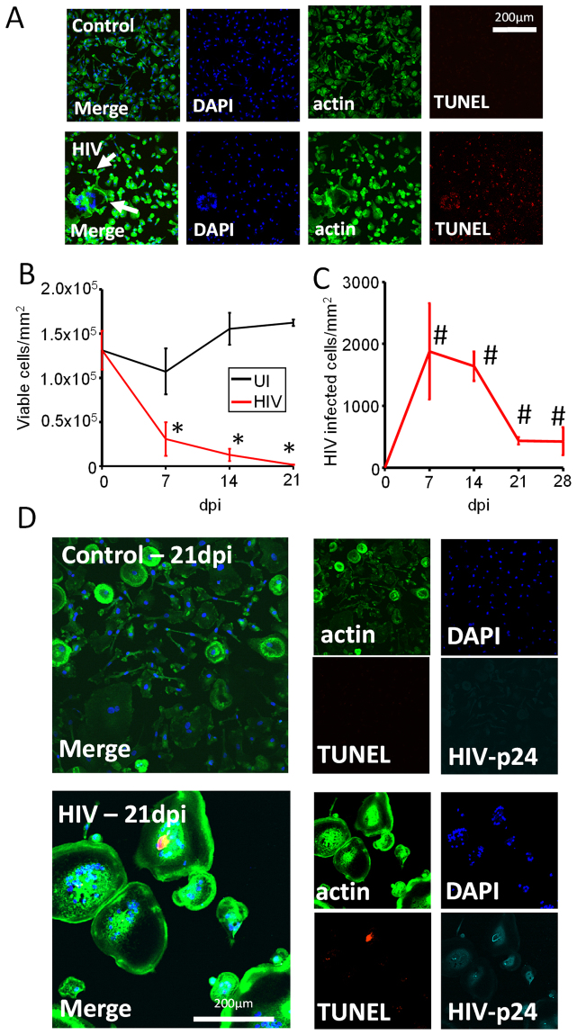
HIV infection of human primary macrophages results in massive apoptosis and survival of a small population of latently infected macrophages. Macrophages were isolated using the same methods as in Fig. 2, and cells were fixed at 7, 14, and 21 days post infection. Cells were stained to identify nuclei (DAPI, blue staining), cell shape (actin/phalloidin, green staining), and apoptosis (TUNEL, red staining). TUNEL positive macrophages were not counted as viable, with exceptions for cases of multinucleated cells. (A) A representative example of apoptosis in control and HIV-infected conditions after 7 days post infection. Arrows denote the presence of fused cells. (B) Quantification of surviving macrophages in control uninfected (UI) or HIV treated cultures (HIV) over 21 days post infection using microscopy. (C) Quantification of surviving HIV-infected macrophages. A significant number of HIV-infected macrophages were present at 7 and 21 days post-infection (#p ≤ 0.0367, n = 3 patients). (D) A representative example of control and HIV-infected cultures at 21 days post infection stained for nuclei (DAPI, blue), cytoskeleton (actin, phalloidin, green), apoptosis (TUNEL, red) and HIV-p24 (Cyan).

