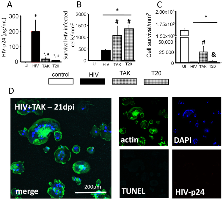
Figure 5.
Fusion inhibitors decreased HIV replication and increased the survival of latentlyHIV infected macrophages. To assess the survival of HIV-infected macrophages in the presence of fusion inhibitors, macrophages were infected with HIVADA, and fusion inhibitors were applied 24–48 hours later. This allowed for infection to establish before application of fusion inhibitors to determine whether fusion inhibitors were useful for inhibiting the formation of MGCs. The supernatant was also collected for application with ELISA. (A) ELISA for HIV-p24 at 21 days post infection in the presence of fusion inhibitors TAK779 (TAK), T20 or HIV alone. Fusion inhibitors TAK and T20 reduced HIV-p24 production collected from the supernatant (#p ≤ 0.0148 as compared to HIV alone, n = 3). Control cultures did not produce an ELISA signal above background (UI). Supernatant from HIV alone cultures contained 196.5 ± 76 pg/mL HIV-p24, a significant increase from control cultures (*p = 0.0112, n = 3). Supernatant collected from HIV infected cultures treated with TAK or T20 also contained a significant amount of HIV-p24 compared with control cultures, 16 ± 8 pg/mL, and 5 ± 2 pg/mL respectively (*p ≤ 0.0123, n = 3). The amount of HIV-p24 in the supernatant of infected cultures treated with TAK or T20 did not significantly differ from each other. The amount of HIV-p24 in supernatant from HIV alone cultures was significantly higher than both TAK779 and T20 treated cultures (#p ≤ 0.015, n = 3). (B) Fusion inhibitors prevented apoptosis of fused HIV-infected macrophages. HIV-infected cultures had significant cell death as compared to UI cultures (*p ≤ 0.0018, n = 3). Cultures treated with TAK779 or T20 contained more HIV-infected macrophages than HIV-alone cultures (#p = 0.0467 as compared to HIV alone, n = 3), indicating TAK and T20 treatment prevented cell death of HIV-infected macrophages over 21 days post infection, up to 28 days the last point assayed. (C) Quantification of non-fused cells reveals that TAK treatment improved survival of non-fused macrophages (most uninfected cells) after exposure to HIV (#p ≤ 0.0341; &p ≤ 0.0428). T20 had no effect compared to HIV-alone cultures (n = 3). (D) An example of cultures treated with TAK779 (TAK) for 24 h and subsequently infected with HIVADA to demonstrate the lack of staining for HIV-p24, significant multinucleation, and cell death as compared to Fig. 3D.

