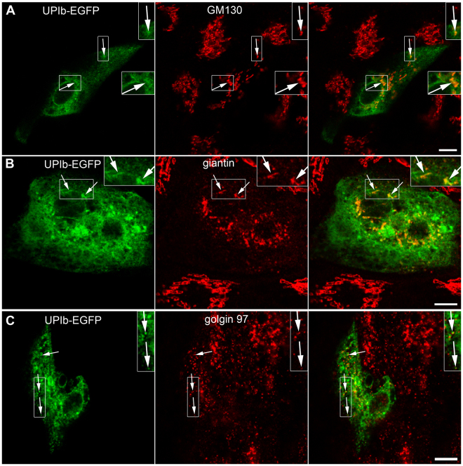
Figure 3.
Expression and localization of UPIb-EGFP in non-polarized UCs. Immunofluorescence labelling of (A) GM130 (cis-GA marker); (B) giantin (cis- and medial GA marker) and (C) golgin-97 (trans-GA network marker) in UPIb-EGFP expressing UCs shows the presence of UPs in different parts of the GA. (A–C) Arrows show the green signals (UPIb-EGFP), red signals (GM130, giantin, golgin 97) and overlap of green and red signals (merged). Areas in larger frames are 50% increased smaller frames. Bars: 10 µm (A–C).

