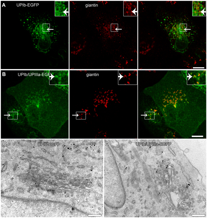
Figure 4.
Expression and localization of UPIb-EGFP and UPIb/UPIIIa-EGFP in non-polarized MDCK cells. MDCK cells were transfected with (A) UPIb-EGFP or (B) co-transfected with UPIb/UPIIIa-EGFP and labelled with the antibody against giantin. (A,B) Both, UPIb-EGFP and UPIb/UPIIIa-EGFP signals overlap with the GA marker giantin (arrows). Smaller frames are 100% enlarged insets on the right. (C,D) The presence of UPIb-EGFP (black dots in C) and UPIb/UPIIIa-EGFP (black dots in D) in the GA is seen also at the ultrastructural level. Bars: 10 µm (A,B); 500 nm (C); 1 µm (D).

