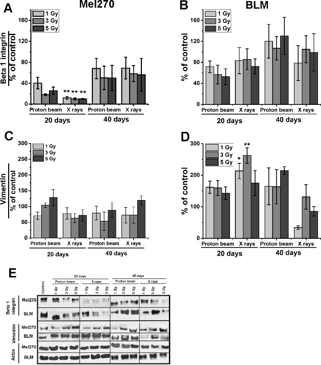
Fig 5.
Integrin β1 (A, B) and vimentin (C, D) protein expression assessed with Western Blot in Mel270 (A, C) and BLM (B, D) cells treated with different doses (1, 3, 5 Gy) of proton beam or X rays. Cells were lysed 20 and 40 days after irradiation. (E) 20 μg of protein was applied per well. Control of untreated cells was set to a 100%. Chemiluminescent evaluation of 3 independent Western blots of cell lysates was shown as a mean of the percentage of the control and SEM. *p<0.05; **p<0.01; ***p<0.001.

