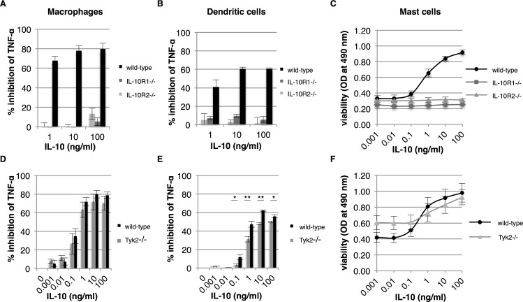
Fig 3. IL-10R2 mediated signaling via Tyk2 plays a limited role in IL-10 activity.
Bone marrow-derived macrophages, dendritic cells and mast cells from wild-type, IL-10R1-/-, IL-10R2-/- and Tyk2-/- mice were tested for their response to IL-10. Macrophages and dendritic cells from wild-type and IL-10R-/- mice were pre-treated with IL-10 and subsequently stimulated with 100 ng/ml LPS. The percentage of inhibition of TNF-α expression of macrophages and dendritic cells was determined after overnight incubation (A and B, respectively) (n = 3, error bars indicate standard error). Similarly, macrophages and dendritic cells from Tyk2-/- mice were tested for their response to IL-10 (D and E, respectively) (n = 4, error bars indicate standard error). Mast cells from wild-type and transgenic mice were cultured for 48 hours in the presence of IL-10 and cell viability was determined (C and F) (n = 3 for IL-10R-/- mice and n = 4 for Tyk2-/- mice, error bars indicate standard error). Asterisk(s) indicate significant differences as determined by a Welch’s t-test (*P<0.05; **P<0.01).

