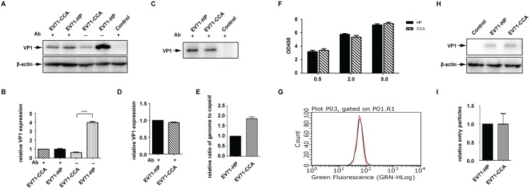
Fig 4. The VP1107 residue modulates viral spread in cell culture.
Equal amounts of infectious RNA of EV71-HP and EV71-CCA were transfected into Vero cells, then treated with EV71 neutralizing antibody 2 hours post transfection. (A) Expression of viral protein in cells treated with the EV71 neutralizing antibody. Cell lysates were subjected to western blot analysis using the antibody specific for EV71 VP1. (B) The band density of each lane shown in panel A was quantified with ImageJ and expressed as fold increase relative to that of EV71-CCA that was treated with the antibody. β-actin was included as the internal control. Three asterisks indicate P<0.01. (C) Viral production from cells treated with the neutralizing antibody. Viral particles in supernatants were collected and purified by ultracentrifugation through a sucrose cushion, and analyzed in western blotting. (D) The band density in panel C was determined with ImageJ and expressed as fold increase relative to band density of EV71-HP. (E) Effect of VP1107 substitution on viral RNA packaging efficiency. The genomic RNA copies were determined by real time PCR, the amounts of capsid protein were determined by Bradford assay. The efficiency of viral RNA packaging is calculated as the ratio of RNA copies to capsid protein. The ratio of EV71-CCA was expressed as fold increase relative to that of EV71-HP. (F) Effect of VP1107 substitution on virus attachment as measured by cell-ELISA. Vero cells were inoculated with virus at different MOIs. The attached viruses were stained withEV71 VP1 antibody and HRP-conjugated secondary antibody. TMB peroxidase substrate was added and the absorbance at OD 450 nm was measured. (G) Effect of VP1107 substitution on virus attachment as measured by flow cytometry. The stained cells shown in panel A were detected by flow cytometer. The red and black curves indicate cells that were infected with EV71-HP and EV71-CCA, respectively. (H) Effect of VP1107 substitution on virus internalization. Vero cells were inoculated with equal amounts of EV71-HP or EV71-CCA. After synchronized adsorption at 4°C for 1 hour, the unbound virions were removed by PBS wash. Virus internalization was performed by incubation at 37°C for 30 minutes. Subtilisin A was used to remove the non-internalized virions. The internalized virions were detected by western blotting. (I) The band density of VP1 of internalized virions shown in panel H was quantified with ImageJ, and expressed as fold increase relative to that of EV71-HP.

