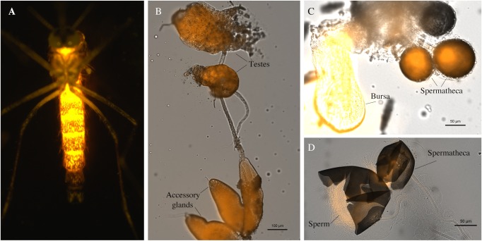
Fig 2. Rhodamine B marking of male seminal fluid.
(A) Example of rhodamine B fluorescence through cuticle of the abdomen of a male Ae. aegypti. (B) Rhodamine B labelling of the testes and accessory glands of male Ae. aegypti. (C) Rhodamine B labelled seminal fluid in the spermathecae and bursa of an Ae. aegypti female transferred during mating with a marked male. (D) Enlarged view of rhodamine B labelled seminal fluid in the spermathecae of an Ae. aegypti that have been opened to reveal the presence of sperm. Males in images A and B were labelled by rhodamine B feeding on a 0.2% w/v solution for 4 days and in image C and D were labelled by rhodamine feeding on a 0.4% w/v solution for 4 days.

