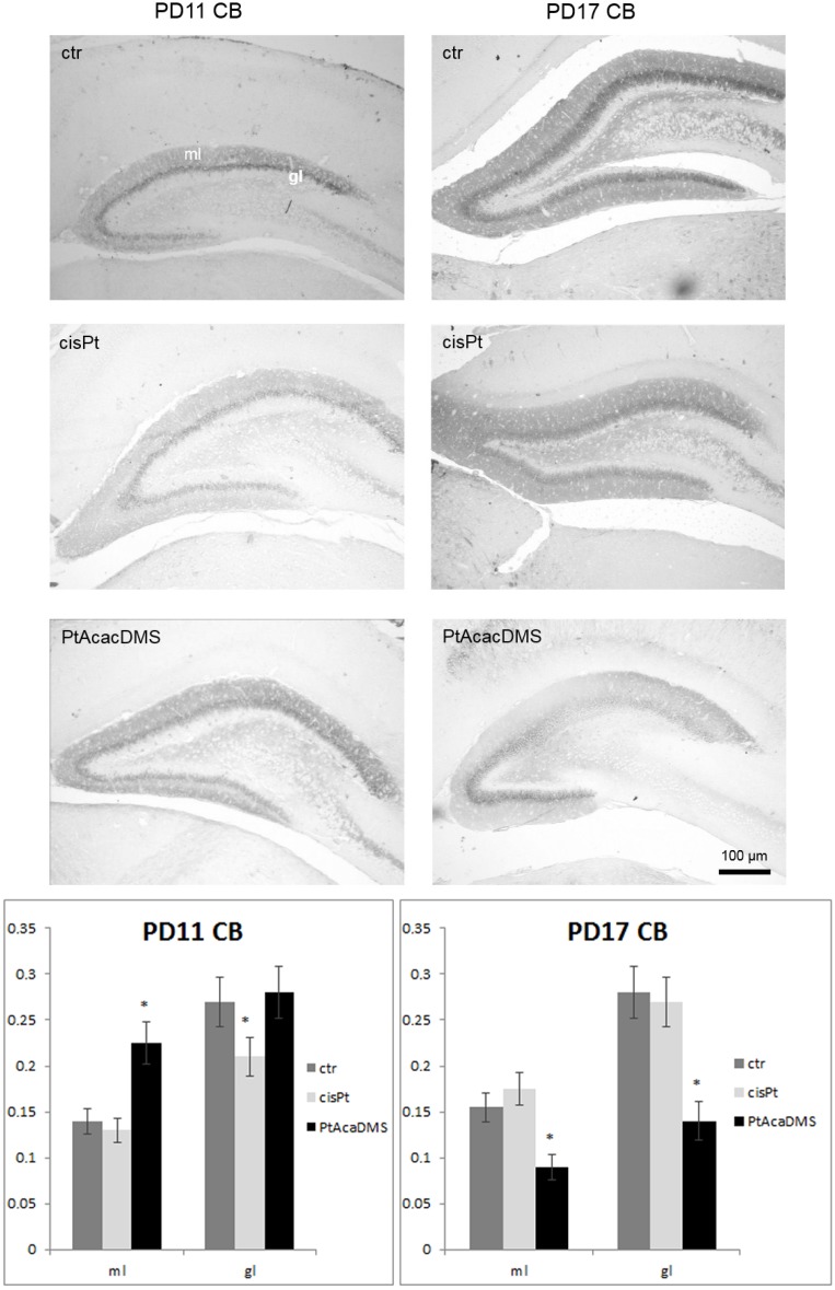
Figure 2.
Calbindin immunocytochemistry in the DG granule cell layer. At PD11, the intensity of labelling in the DG granule cells decreases after cisPt and increases after PtAcacDMS. At PD17, decreased immunolabelling intensity is found after PtAcacDMS and not after cisPt. Histograms show the OD values and the significance of differences is reported (* p < 0.05). ml: Molecular layer; gl: Granule cell layer. Scale bar: 100 μm.

