. 2017 Sep 25;6:e28199. doi: 10.7554/eLife.28199

Figure 2. The absence of SUN5 has no effect on acrosome biogenesis but disrupts the development of the coupling apparatus between sperm head and tail.

(A) IF (immunofluorescence) staining of sp56 in WT and Sun5-null spermatozoon. The Sun5-null spermatozoa contains both round-headed spermatozoa and tailless heads (lower two panels). The proportion of these two types of spermatozoa were displayed in Figure 1J. Note that the round-headed Sun5-null spermatozoa do not contain nuclei and acrosomes, but the tailless Sun5-null sperm heads have nuclei and acrosomes. Scale bar: 5 μm. (B) Ultrastructure of WT and Sun5−/−caudal epididymides showing that the Sun5-null spermatozoon was filled with cytoplasm and misarranged mitochondria. Note that the axoneme of Sun5-null spermatozoon was also disrupted. Scale bar: left panel, 1 μm; right panel, 200 nm. (C) TEM analyses of the stepwise development of the coupling apparatus in WT and Sun5-null spermatozoa. In the round spermatid stage, the coupling apparatus can be assembled in both WT and Sun5-null spermatid, but the coupling apparatus could not be tightly attached to the nuclear envelope in Sun5-null spermatids. The asterisk indicates the gap between the nuclear (Nu) envelope and the basal plate (Bp). In the following developmental stages, the coupling apparatus was well-fixed on the nuclear envelope in WT spermatids, ensuring healthy spermatid differentiation. While in Sun5-null spermatids, the basal plate (Bp)-capitulum (Cp)-segmented column (Sc) together with the centriole (Pc) was detached from the nuclear envelope during spermatid elongation. An, annulus. Scale bar: the 1st and 3rd panel, 2 μm, 2nd and 4th panel, 0.5 μm.

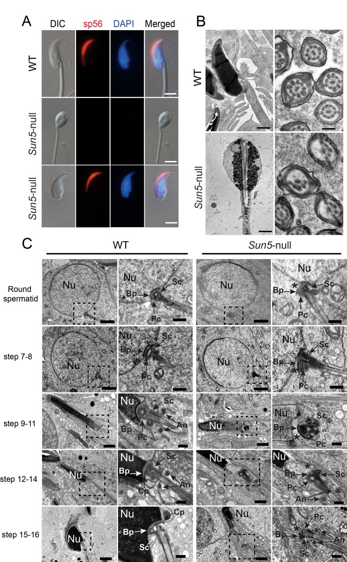
Figure 2.

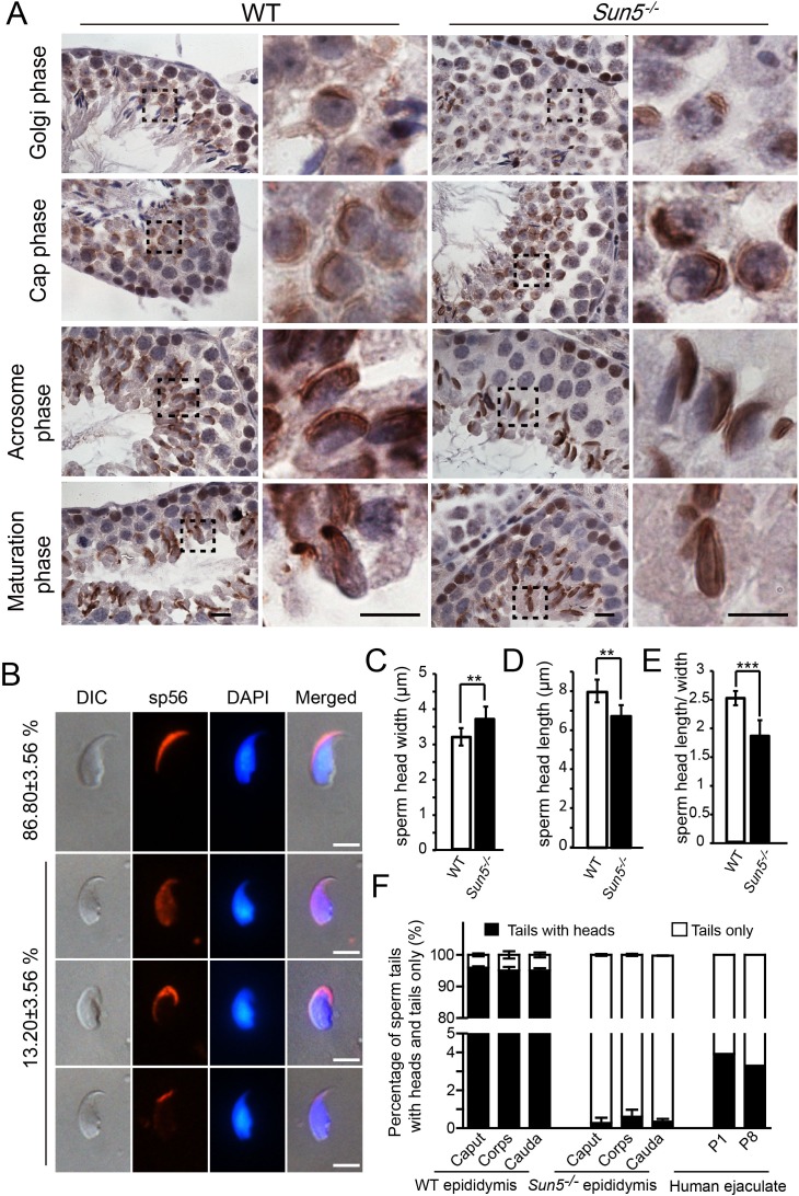
Figure 2—figure supplement 1. Acrosome biogenesis and epididymal spermatozoa in Sun5−/− testes.

Figure 2—figure supplement 1.

(A) Immunohistochemistry staining of Afaf in WT and Sun5−/− testes, indicating that acrosome biogenesis was not affected in Sun5−/− testes. Scale bar: 10 μm. (B) sp56 IF staining in Sun5–null spermatozoa, showing that most Sun5–null spermatozoa had normal acrosome. Scale bar: 5 μm. (C) The width of sperm heads in WT and Sun5–null spermatozoa. (D) The length of sperm heads in WT and Sun5–null spermatozoa. (E) The ratio of sperm head length to width in WT and Sun5–null spermatozoa. Showing the differences between WT and Sun5–null sperm head morphology. (F) The percentage of spermatozoa with heads versus tails only in mouse caput, corpus, cauda epididymis and human ejaculate, and very few intact spermatozoa could be found in Sun5 mutant mouse and human.
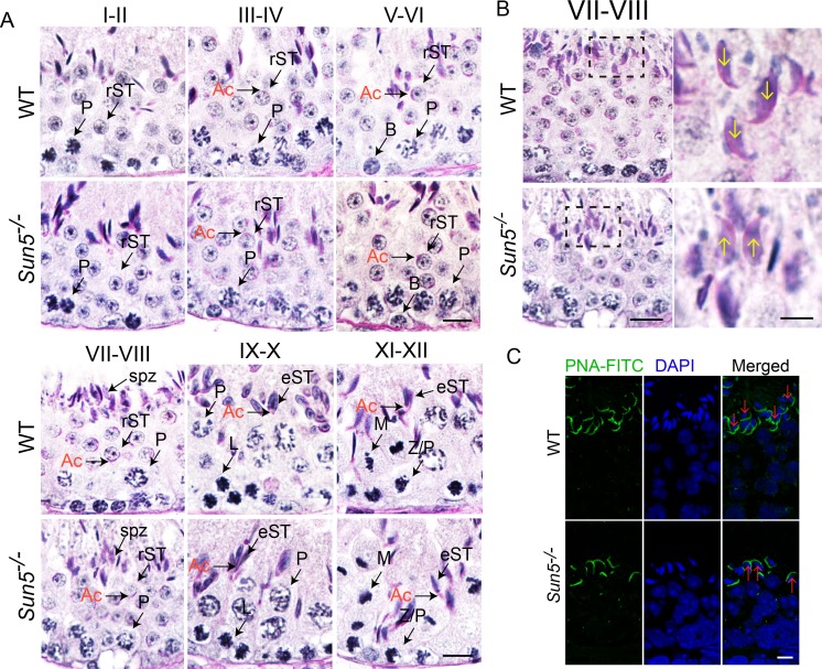
Figure 2—figure supplement 2. Spermiation defects in Sun5−/− mice.

Figure 2—figure supplement 2.

(A) Periodic acid-Schiff (PAS) staining revealed histology of all twelve developmental stages in WT and Sun5−/− testes. The morphology of Acrosome (Ac) marks the specific developmental stages. Scale bar, 10 μm. (B) Sun5-null spermatids have lost their orientation toward the basement membrane during spermiation in stage VII-VIII seminiferous epithelia. The arrows indicate the orientation of the sperm heads. Scale bar: left panel, 10 μm; right panel, 5 μm. (C) Peanut agglutinin (PNA) staining of stage VII-VIII seminiferous epithelia showed the miss-arranged spermatids in Sun5−/− testes. The arrows indicate the orientation of the sperm heads. Scale bar, 5 μm. Ac: acrosome; B: B type spermatogonium; eST: elongating spermatid; L: leptotene spermatocyte; M: meiotic spermatocyte; P: pachytene spermatocyte; rST: round spermatid; spz: spermatozoon; Z: zygotene spermatocyte.