
Fig. 6.
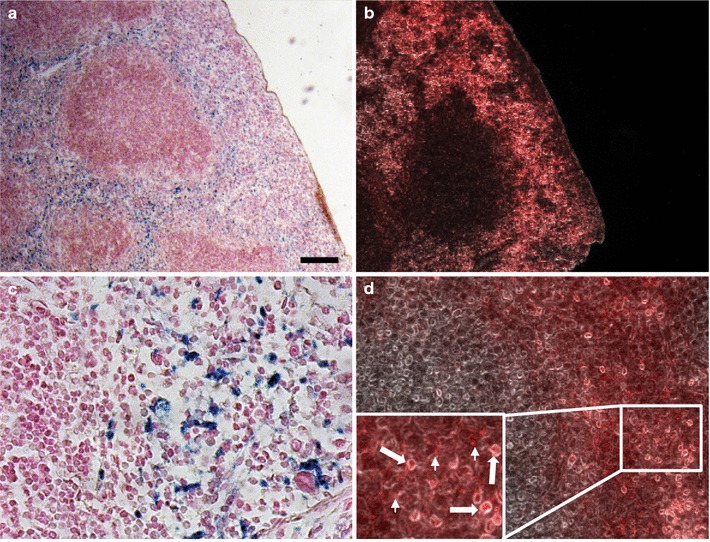
Fluorescence detection of Eu-VSOP in tissue sections. Ex vivo detection of Eu-VSOP in mouse spleen. 24 h after IV administration of Eu-VSOP, iron (blue) was detected in splenic red pulp (a, c, Prussian blue stain with nuclear fast red counterstain). Serial section with fluorescence detection of europium with HEE (b, d, red staining, bright-field background). Inset in d shows intracellular and extracellular Eu-VSOP accumulations (arrows and arrowheads, respectively). Scale bar: 100 µm