Fig. 3.

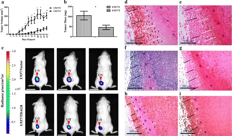
Restoring TDAG8 gene expression in U937 cells reduces primary tumor growth in SCID mice. a U937/TDAG8 tumor growth was significantly reduced in comparison to U937/Vector tumors from day 4 to necropsy (N = 14). b U937/TDAG8 tumor mass was reduced significantly in comparison to U937/Vector tumors following necropsy. c In vivo imaging of 3 mice injected with U937/Vector-Luc or U937/TDAG8-Luc cells 6 days after injection. Scale Bar = 2 cm. d, e Immunohistochemistry of c-myc d and cleaved PARP e in U937/Vector tumors demonstrating regions nearest necrotic areas display reduced c-myc oncogene expression while still live. f–i Hematoxylin and eosin staining (f) and immunohistochemistry of U937/Vector tumors demonstrating invasive peripheral regions of U937 tumors display increased proliferation by c-myc (h) and Ki67 (i) while demonstrating less apoptosis, cleaved PARP (g). N necrotic. White lines indicate areas adjacent to necrotic zones. Black lines indicate tumor cells that are invasive correlating with higher c-myc and Ki67 expression. Black arrowheads indicate single tumor cells that demonstrate reduced or no c-myc expression. *P < 0.05, **P < 0.01, ***P < 0.001
