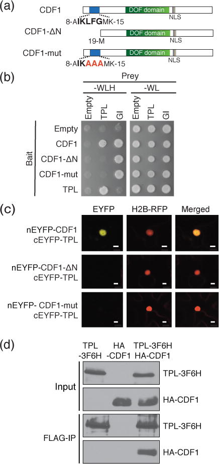
Figure 1. CDF1 and TPL form a protein complex through a conserved binding motif located at the N-terminus of CDF1.

(a) Schematic representation of CDF1 full length protein and the N-terminal CDF variants used in this study. The N-terminal amino acid sequences (IKLFG) conserved in CDF family proteins (Figure S1), which overlap with the TPL binding motif (R/KLFGV), are shown in bold. The mutated amino acids in CDF1-mut protein are shown in red. The relative positions of DOF DNA binding domain (DOF domain) and nuclear localization signal (NLS) are indicated. (b) Y2H analysis of CDF1-TPL protein interaction. The –WLH plate shows the interaction of bait and prey proteins, while the –WL plate shows the presence of both bait and prey constructs. N-terminus of GI protein was used as a positive control for CDF1 variants. (c) BiFC interaction analysis in transiently transfected N. benthamiana leaves between full-length of CDF1 protein, CDF1-ΔN and CDF1-mut variants, and TPL protein. Histone H2B-RFP was used to determine the position of the nucleus in the same cell. Scale bars show 10 μm. (d) TPL-CDF1 interaction using coimmunoprecipitation assay of transiently transfected N. benthamiana leaves.
