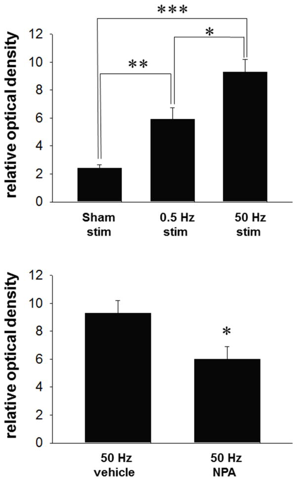
Figure 4. Increases in NADPH-d staining elicited in the msNAc via fimbria stimulation is dependent on frequency of electrical stimulation and nNOS activity.
Top: Low frequency stimulation (0.5 Hz for 20 minutes duration) increased staining in the msNAc compared to sham simulated control animals (electrode implanted with no stimulation) (**p<0.01). High frequency stimulation (50 Hz at 750μA, 0.5 ms, 2 s ITI for 20 minutes duration) significantly increased NADPH-d staining in the msNAc compared to both sham simulated controls (***p<0.001) and animals receiving low frequency (0.5 Hz) stimulation (*p<0.05). Results are expressed as the mean ± S.E.M. from n=5–6 rats per group. Bottom: Staining elicited by high frequency stimulation (50 Hz at 750μA, 0.5 ms, 2 s ITI for 20 minutes duration) was significantly reduced in the msNAc after NPA administration (*p<0.05). Results are expressed as the mean ± SEM from n= 5 rats per group.

