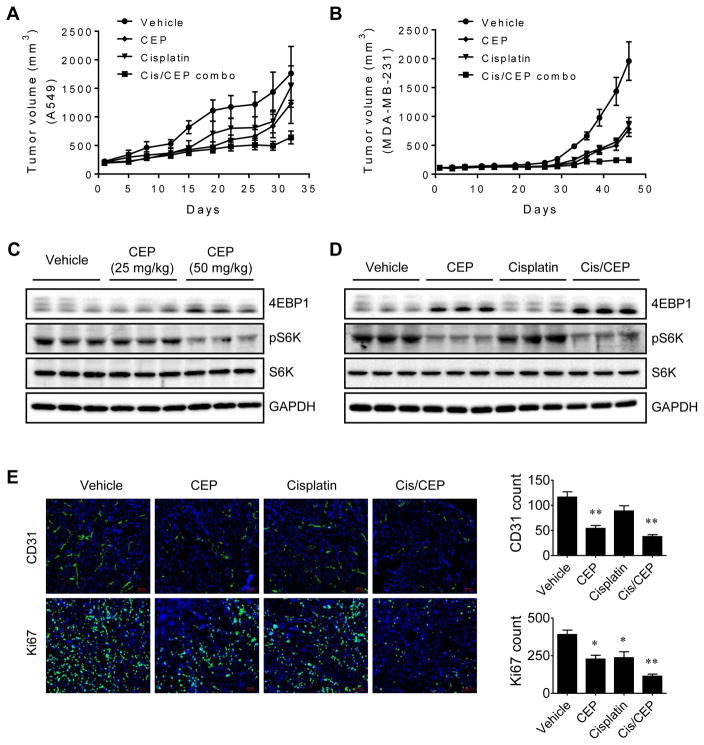
Figure 7.
Antitumor and antitumor-promoting effect of CEP on lung and breast cancer xenografts. (A and B) Tumor volume measurements of A549 lung cancer (A) and MDA-MB-231 breast cancer (B) xenografts in mice are shown. CEP (50 mg/kg, daily), cisplatin (5 mg/kg, weekly), or combination of CEP and cisplatin (Cis/CEP combo) was administered intraperitoneally into the xenograft mice. (C and D) The tumor tissues from mice were isolated and analyzed with Western blots for 4EBP1 and S6K phosphorylation to assess the mTOR down-stream signaling in the tumor. (E) Tumor tissues were processed for immunofluorescence staining of blood vessel marker, CD31 and cell proliferation marker, Ki67. Representative immunofluorescence images are shown on the left. Scale bar = 100 μm. The number of blood vessels and Ki67-positive tumor cells were quantified and shown on the right. *P < .05 and **P < .01 vs vehicle control.

