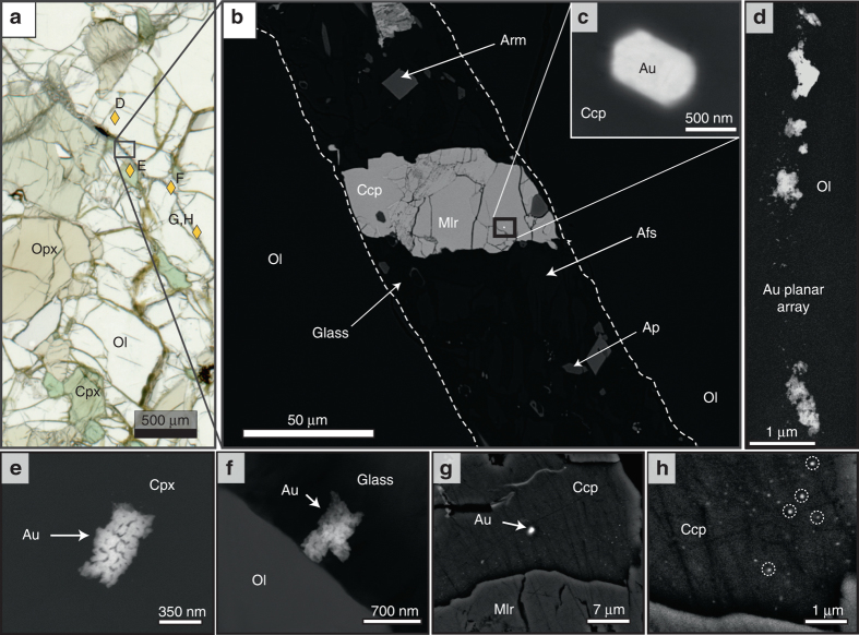
Fig. 2.
Photomicrograph and backscattered electron (BSE) images of Au particles in the Cerro Redondo mantle xenolith. a Plane polarised light image of the lherzolite sample showing the late metasomatic glass vein and the location of Au particles (golden diamonds and letters refer to BSE images). b–h Backscattered electron FE-SEM images of Au particles and their microstructural setting. b Detail of the glass vein showing its metasomatic assemblage and a composite sulfide grain containing a Au particle. c Magnification of the euhedral Au particle within chalcopyrite from the composite sulfide grain in b. d Planar array of Au particles within olivine. e Au particle enclosed within clinopyroxene. f Au particle within the glass of the metasomatic vein in contact with olivine. g Au particle within chalcopyrite and arrangement of Au nanoparticles enlarged in h. Afs, alkali feldspar; Ap, apatite; Arm, armalcolite; Ccp, chalcopyrite; Cpx, clinopyroxene; Mlr, millerite; Ol, olivine; Opx, orthopyroxene

