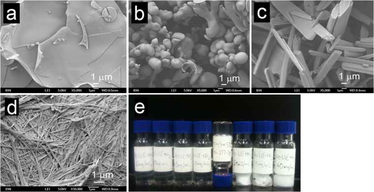
Figure 5.
Scanning electron micrographs illustrating the four general morphologies observed with the acetylated tripeptides: (a) Amorphous structure (Ac-LVE, supernatant of 20 mg/mL; 5000×). (b) Bead microstructure (Ac-LLE, solution of 20 mg/mL; 5000×). (c) Crystalline nanostructure (Ac-LVE, precipitate of 20 mg/mL; 5000×). (d) Fibrillar nanostructure (Ac-IVD, hydrogel at 20 mg/mL; 10000×). (e) Illustration of a typical set-up to assess the self-assembly and aggregation of peptides. A series of peptide concentrations (Ac-LLE-NH2 here) from 5–40 mg/mL, in steps of 5 mg/mL were prepared. This series also illustrates the four states generally observed in this study: solution (5–20 mg/mL), hydrogel (25 mg/mL; upturned vial), supernatant and precipitate (30–40 mg/mL).

