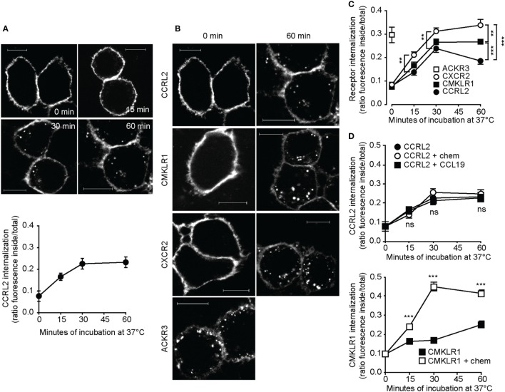
Figure 1.
C-C chemokine receptor-like 2 (CCRL2) internalization is ligand independent. HeLa cells were transfected with acyl carrier protein (ACP)-CCRL2, ACP chemokine like receptor 1 (CMKLR1), peptidyl carrier protein-C-X-C motif chemokine receptor 2 (PCP-CXCR2), or PCP-atypical chemokine receptor 3 (ACKR3) expression vectors and receptors were labeled. Cells were then incubated at 37°C in serum-free medium for the indicated times and fixed. Images were taken using 100× magnification and analyzed using ImageJ 1.48 (NIH). (A) Representative images (above) and quantification (below) of ACP-CCRL2 internalization after 0, 15, 30, 60 min of incubation. Internalization is expressed as the ratio between internal and total fluorescence, calculated on each single cell. One experiment representative of two is shown; each point represents the average value ± SEM calculated on 19–32 cells. (B) Representative images of labeled ACP-CCRL2, ACP-CMKLR1, PCP-CXCR2, and PCP-ACKR3, at 0 and 60 min incubation. Scale bars: 10 µm. (C) ACP-CCRL2, ACP-CMKLR1, PCP-CXCR2, and PCP-ACKR3 internalization, calculated as in (A). One experiment representative of two is shown; each point represents the average value ± SEM calculated on 20–50 cells. **p < 0.01 and ***p < 0.001 by two-way ANOVA. (D) (Above) After labeling of ACP-CCRL2, cells were incubated in the presence of 100 nM CCL19 or 100 nM chemerin for the indicated times. Internalization was measured as described before; analysis by two-way ANOVA; ns, not significant (CCRL2 vs. CCRL2 + chem and CCRL2 + CCL19). (Below) After labeling of ACP-CMKLR1, cells were incubated in the presence or in the absence of 100 nM chemerin for the indicated times; internalization was measured as described before. ***p < 0.001 by two-way ANOVA (CMKLR1 vs. CMKLR1 + chem). In both the graphs, one representative of two independent experiments is shown, each point represents the average values ± SEM calculated on 20–50 cells.

