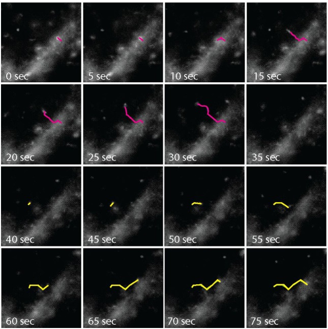
Figure 2.

Time-lapse microscopy analysis of C-C chemokine receptor-like 2 (CCRL2)-positive vesicles. COS-7 cells expressing acyl carrier protein (ACP)-CCRL2 were enzymatically labeled. After labeling, cells were placed under the microscope at 37°C, in the presence of 5% CO2, and observed by time-lapse microscopy every 5 s. Pink line, endocytosing vesicle; yellow line, exocytosing vesicle.
