Figure 3.
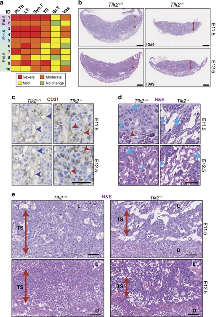
TLK2 is required for placental development. (a) Semiquantitative analysis of histological phenotypes in Tlk2−/− placentas. In each case, Tlk2−/− placentas (n=10) at the indicated age were compared with wild-type littermates. Phenotypes are abbreviated as follows: placental thickness (Pl.Th), numbers/size of Labyrinth trophoblasts (LT), syncytiotrophoblasts (Syn.T), spongiotrophoblasts including glycogen cells (TS), giant cell trophoblasts (Gi.T) and vasculature (Vas). Summary of the grading system used48 was provided in the Materials and Methods. (b) Sections from E11.5 and E12.5 littermate placentas stained with Hematoxylin and eosin (H&E). Tlk2−/− placentas are thinner, less cellular and poorly vascularized compared with Tlk2+/+. Scale bars=500 μm. (c) CD31 staining reveals that the vasculature of Tlk2−/− placentas is miniaturized, collapsed and appears slit-like (red arrowheads) compared with the well-developed, widely opened vasculature of Tlk2+/+ (blue arrowheads). Scale bar=20 μm. (d) H&E staining from the indicated age and genotype showing fewer and smaller trophoblasts (arrows) and syncytiotrophoblasts (blue arrowheads) with an absence of fetal vasculature (red arrowhead) in Tlk2−/− placentas. Scale bar=20 μm. (e) H&E staining of placentas showing a thinner trophospongium (spongiotrophoblasts and glycogen cells) in Tlk2−/− placentas. The trophospongium (TS), labyrinth (L) and decidua (D) are indicated. Scale bar=50 μm

