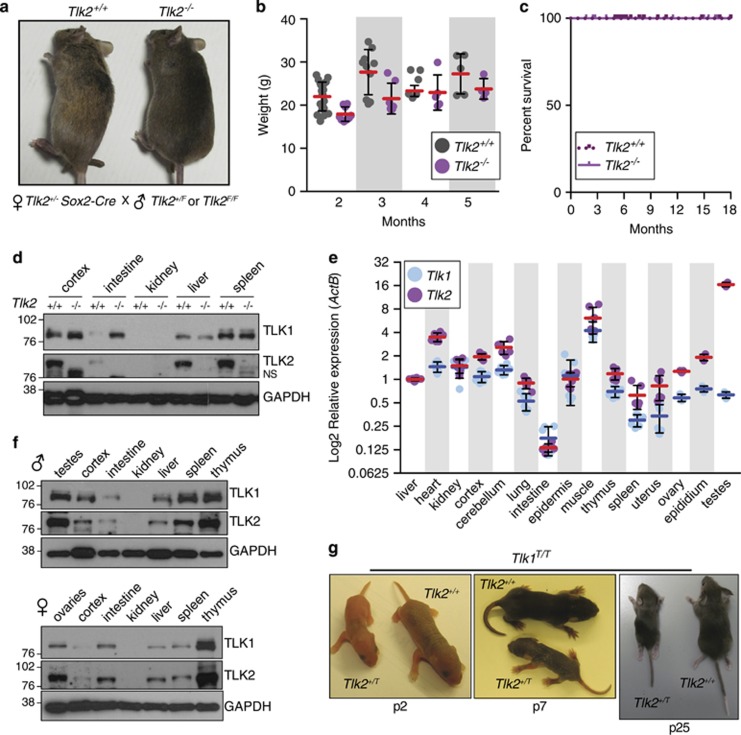
Figure 6.
TLK2 is dispensable in embryonic and adult tissues. (a) Tlk2+/+ and Tlk2−/− littermates are shown from breedings of female Tlk2+/−, Sox2-Cre+ and male Tlk2+/F mice. (b) Weight of mice lacking TLK2 is slightly reduced but growth is similar to wild type. Mean (red bars) and S.D. are indicated (n=20 and 9 for 1 month, n=10 and 7 for 2 months, n=6 and 5 for 3 months, n=5 and 4 for 4 months for Tlk2+/+ and Tlk2−/−, respectively). (c) Kaplan–Meier survival curve of mice with the indicated genotypes (Tlk2+/−, n=42; Tlk2−/−, n=26). (d) Western blotting of TLK1 and TLK2 using lysates from selected tissues of Tlk2+/+ and Tlk2−/− mice. NS refers to a nonspecific band observed with the TLK2 antibody in many tissues and cell lines. (e) Similar expression patterns of Tlk1 and Tlk2 in adult mice. Taqman real-time PCR analysis of Tlk1 and Tlk2 mRNA levels (log 2-transformed) relative to ActB. Mean (blue bars for Tlk1 and red bars for Tlk2) and S.D. of triplicate reactions from at least two animals are plotted. In each case, levels are normalized to that of the liver (set to 1). (f) Western blotting of TLK1/2 protein levels in 2-month-old male and female wild-type tissues. Glyceraldehyde 3-phosphate dehydrogenase (GAPDH) is shown as a loading control. (g) Examples of littermate Tlk1T/T Tlk2+/+ and Tlk1T/T Tlk2+/T animals at different ages. All Tlk1T/T Tlk2+/T animals (n=4) observed were severely runted and one lacked limbs on the left torso. All mice were killed before 1 month following veterinary advice owing to the lack of mobility and trembling. A Kaplan–Meier survival plot for all related genotypes is presented in Supplementary Figure S7

