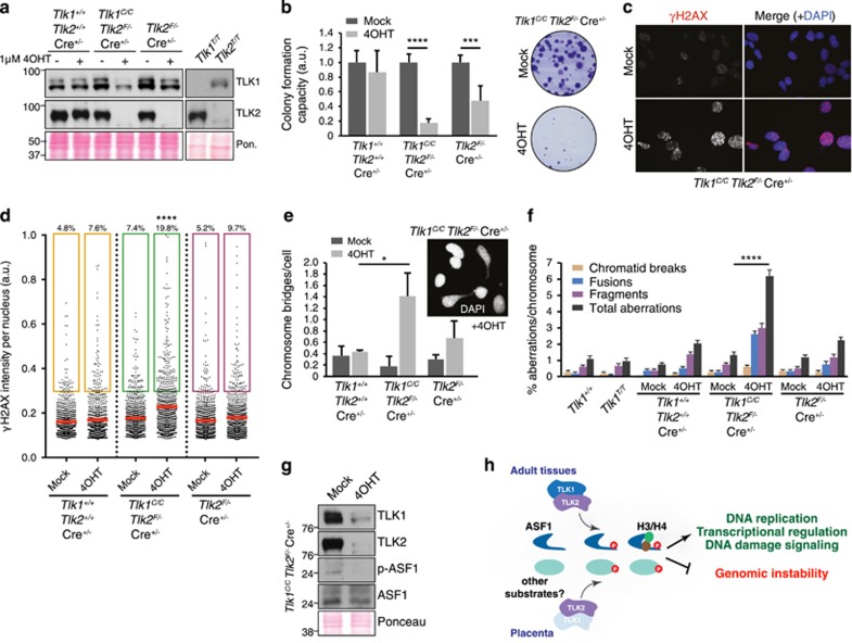
Figure 7.
TLK1 and TLK2 cooperate to maintain cell viability and chromosome stability. (a) Western blot with antibodies against TLK1 and TLK2 in whole-cell extracts of transformed MEFs of the indicated genotypes mock-treated or treated with 4OHT to induce trapping of TLK1 and/or deletion of TLK2 (Pon.=Ponceau red-stained membrane). (b) Deletion of TLKs reduces colony formation. MEFs were mock-treated or treated with 4OHT for 72 h, washed, plated and cultured for 10–14 days. Relative colony number to mock-treated cells is displayed. Tlk1+/+ Tlk2+/+ Cre+/−, n= 3; Tlk1C/CTlk2F/− Cre+/−, n=6; Tlk2F/− Cre+/−, n=6. Results are shown as mean±S.E.M. Experiment was performed in technical duplicates, at least in biological triplicate. Statistical significance was determined using an upaired t-test (****P<0.0001, ***P<0.001). (c) Representative images of IF experiments staining for γH2AX are shown from Tlk1T/T Tlk2F/− cells that were mock-treated or treated with 4OHT. Nuclei were counterstained with 4′,6-diamidino-2-phenylindole (DAPI). (d) Increased DNA DSB formation following the depletion of TLK1 and TLK2. High-throughput microscopy of γH2AX levels per individual nucleus in response to TLK depletion in MEFs mock-treated or treated with 4OHT. At least 800 nuclei were quantified per condition. Mean is shown as a red line; a.u., arbitrary units. Statistical significance was determined using ordinary one-way analysis of variance (ANOVA) Tukey's multiple comparisons test (****P<0.0001). (e) Increased chromosome bridges are evident in 4OHT induced double TLK-deficient cultures relative to mock-treated or Tlk2 deletion alone. A minimum of 200 cells was scored in each independent experiment. Tlk1+/+ Tlk2+/+ Cre+/−, n= 2; Tlk1C/CTlk2F/− Cre+/−, n=5; Tlk2F/− Cre+/−, n=4. Results are shown as mean±S.D. Statistical significance was determined using an upaired t-test (*P<0.05). (f) Plot of chromosomal aberration types scored from multiple transformed MEF cultures depicted as percent aberrations per chromosome. Results are shown as mean±S.E.M. Statistical significance was determined using Fisher’s exact test (****P<0.0001). (g) Western blotting of Tlk1C/CTlk2F/− MEF lysates mock-treated or treated with 4OHT for TLK1, TLK2, ASF1 and p-ASF1. Experiment performed in biological triplicate and a representative example shown. (h) Proposed model for the contribution of TLK1 and TLK2 activity to the placenta and adult tissues. In the placenta, and possibly other cell types or tissues, TLK2 is the prevalent activity and is required to maintain a threshold of signaling required for viability. This likely involves ASF1a/b and potentially other substrates. In most adult tissues, TLK1 and TLK2 are largely redundant and the presence of either is sufficient to provide the necessary activity to support cellular functions required for the maintenance of genome integrity and viability

