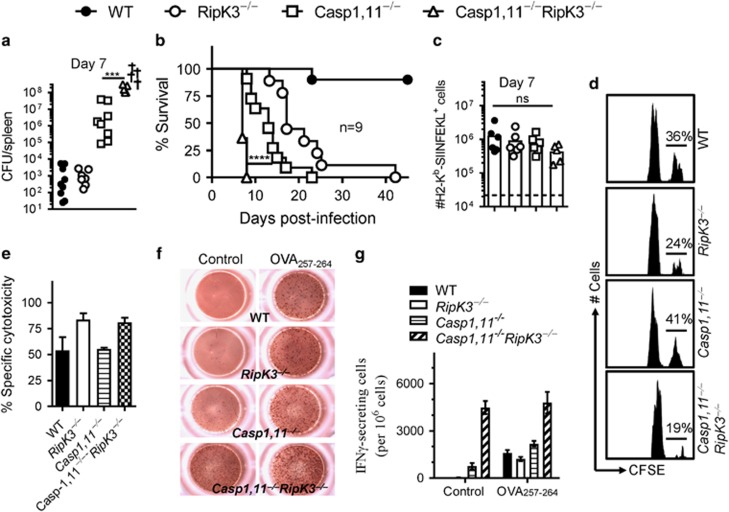
Figure 5.
Synergism of Caspase-1/11 and RipK3 signaling promotes control of ST infection. WT, RipK3−/minus;, Caspase-1,11−/minus; and Caspase-1,11−/minus;RipK3−/minus; mice were infected with ST-OVA (103, i.v.). On day 7, bacterial burden was evaluated in the spleens of infected mice (a). Survival of infected mice was monitored for up to 60 days (b). The numbers of OVA257-264 (SIINFEKL)-specific CD8+ T cells were evaluated in the spleens of infected mice by staining with anti-CD8 antibody and H2-Kb-SIINFEKL tetramers (c). Naive spleen cells (control- CFLElow and OVA257-264-pulsed- CFSEhi) were injected into mice that were infected with ST-OVA 6 days prior. At 24 h post spleen cell transfer, the fate of transferred cells was evaluated in the spleens of infected mice (d,e). Mice were infected as described above, and CD8+ T cells were purified on day 7 post-infection, and the expression of IFN−γ evaluated by ELISPOT assay with/without OVA257-264 pulsed naive spleen cells (f,g). Data is shown as mean ±SEM and is representative of 2–3 separate experiments

