Figure 6.
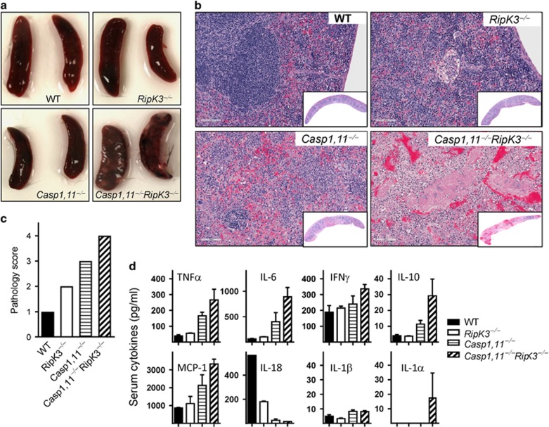
Exacerbated tissue pathology in Caspase-1,11–RipK3-double-deficient mice. WT, RipK3−/minus;, Caspase-1,11−/minus; and Caspase-1,11−/minus;RipK3−/minus; mice were infected with ST-OVA as described in Figure 5. Representative images of spleens harvested from surviving mice at day-7 post-infection with ST-OVA are shown (a). Spleens were collected (day-7 post-infection) and treated with 10% formalin and vertical sections were stained for hematoxylin and eosin (H&E) (b) and the extent of pathology was scored (c). Serum was also collected at day-5 post-infection and cytokine expression measured (d). The data are shown as mean ±SEM and is representative of 2–3 separate experiments

