Fig. 8.
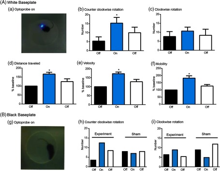
Validation of CerebraLux in vivo. (A) White HDPE. Mice (Ai27 x D1-cre, ) were implanted with the baseplate, made from white HDPE, containing the fiber-optic (the optic component) and allowed to recover. Shortly before the experiment, the PCB (the electronic component) was attached and mouse placed in the open field for 5 min, the probe was turned on for 5 min (a) and then off for the last 5 min. The behavior was video-tracked and the data exported and analyzed. We found that ChR2 activation in the right striatum (b) increased the number of counter-clockwise rotations, (c) did not alter the number of clockwise rotations, and increased distance traveled (% baseline, d), velocity (% baseline, e), and time spent mobile or % mobility (% baseline, f). * versus the first “OFF” period. (B) Black HDPE. Mice (Ai27 x D1-cre, ) were implanted with the baseplate, made from black HDPE, and the same experiment was conducted as in A (experiment). There was a marked reduction in light seen when the probe was turned on (g) but the same relative increase in counter-clockwise, but not clockwise, rotations was observed as in mice implanted with a white HDPE baseplate. A sham mouse (Ai27 × D1 cre), implanted with an optoprobe without the intracerebral fiber-optic, showed no counter-clockwise rotations and a decrease in clockwise rotations when the probe was turned on (h) and (f).

