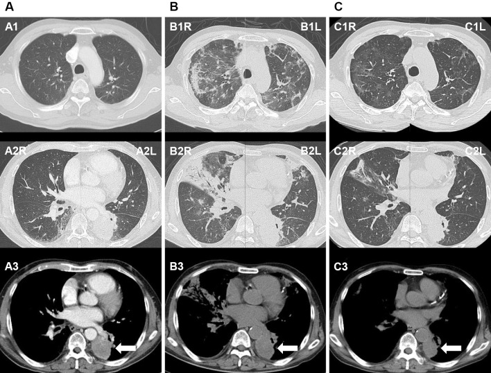
Figure 1.
A: Before osimertinib administration, a solid tumor is observed in the left lower lung (white arrow) with no evidence of interstitial pneumonia. B: After admission, at 34 days after osimertinib initiation, there are diffuse ground glass opacities, areas of patchy consolidation observed in both lungs, and peribronchial consolidation on the right middle lobe. There is no remarkable change in the size of the primary tumor (white arrow). C: Follow-up on the 16th hospital day, after steroid therapy, reveals the resolution of the bilateral ground glass opacities and areas of consolidation, as well as a decrease in the size of the primary tumor (white arrow).

