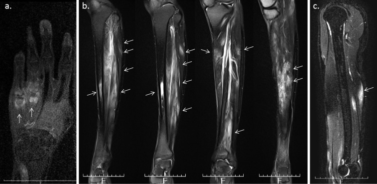
Figure 4.
a: MRI (T1WI) of the right hand reveals synovitis and bone marrow edema of the fourth and fifth metacarpophalangeal joint. b: MRI (T2WI) shows enhanced bone marrow in the right tibial diaphysis associated with cortical bone hypertrophy. The right lower leg muscle was also enhanced, suggesting myositis. c: MRI (T2WI) reveals an enhancement of the right triceps brachii muscle.

