F1000Research logoLink to F1000Research
. 2017 Oct 2;5:2532. Originally published 2016 Oct 18. [Version 2] doi: 10.12688/f1000research.9473.2

Case Report: Whole exome sequencing identifies variation c.2308G>A p.E770K in RAG1 associated with B- T- NK+ severe combined immunodeficiency

Geeta Madathil Govindaraj 1,#, Shamsudheen Karuthedath Vellarikkal 2,3,#, Rijith Jayarajan 3, Rowmika Ravi 3, Ankit Verma 3, Krishnan Chakkiyar 1, Machinari Puthenpurayil Jayakrishnan 1, Riyaz Arakkal 1, Revathi Raj 4, Rajeevan Kunnaruvath 5, Sridhar Sivasubbu 2,3,a, Vinod Scaria 2,6,b
PMCID: PMC5635439  PMID: 29067161

Version Changes

Revised. Amendments from Version 1

This updated version of the manuscript is incorporated with reviewers’ suggestions and more information on clinical findings of patient. A new author, Dr Rajeevan Kunnaruvath, was added to this new version of the manuscript because of his contribution in interpreting the flow cytometry data of the patient.

Abstract

Severe combined immunodeficiency is a large clinically heterogeneous group of disorders caused by a defect in the development of humoral or cellular immune responses. At least 13 genes are known to be involved in the pathophysiology of the disease and the mutation spectrum in SCID has been well documented. Mutations of the recombination-activating genes RAG 1 and RAG 2 are associated with a range of clinical presentations including, severe combined immunodeficiency and autoimmunity. Recently, our understanding of the molecular basis of immune dysfunction in RAG deficiency has improved tremendously with newer insights into the ultrastructure of the RAG complex. In this report, we describe the application of whole exome sequencing for arriving at a molecular diagnosis in a child suffering from B- T- NK+ severe combined immunodeficiency. Apart from making the accurate molecular diagnosis, we also add a genetic variation c.2308G>A p.E770K to the compendium of variations associated with the disease.

Keywords: Severe Combined Immunodeficiency, B- T- NK+ SCID, Whole Exome Sequencing, RAG1

Introduction

Severe combined immunodeficiency (SCID) encompasses a constellation of clinically and genetically heterogeneous diseases resulting in defects of the humoral and/or cellular immune defense mechanism 1. The patients with SCID exhibit recurrent infections with bacteria, virus and fungi. The deficiency of recombination activating gene is associated with T -B -NK + SCID. Recombination of activating gene enzymes plays a significant role in recombination of V(D)J segments 2. The mutation in recombination activating gene 1 ( RAG1) is associated with absence of V(D)J recombination, which in turn produces immature lymphocytes leading to SCID 3. The accurate molecular diagnosis in SCID enables genetic counselling for disease 4. So far, arriving at a precise molecular diagnosis has been quite cumbersome, technically challenging and expensive, as over a dozen genes are known to be implicated in the genetic disease, which would require systematic targeted sequencing of each of the gene 5, 6. The advent of next generation sequencing, especially whole exome and sometimes whole genome sequencing has significantly enabled the rapid identification of the causative genetic variations in clinical settings 6.

In this report, we describe the application of whole exome sequencing for the accurate molecular diagnosis of a case of T -B -NK+ SCID. Our report also adds a genetic variation c.2308G>A p.E770K in Recombination activating gene 1 ( RAG1) to the compendium of variations associated with the disease.

Case report

Here we report a case of a seven-month-old boy, born out of a third degree consanguineous marriage, with a history of recurrent episodes of pneumonia, acute otitis media, diarrhea and oral thrush since two months of age. The child was pale, emaciated, febrile, and had respiratory distress with lower chest retractions. He was in compensated shock. There was no clubbing, cyanosis or lymphadenopathy. There was no facial dysmorphism, and skin and hair were normal. He weighed 5.4 kg; measured 64 cm in length and head circumference was 39.5 cm; all below the 3rd centile as per WHO Child Growth Standards. Examination of the chest showed evidence of bronchopneumonia while there was no evidence of congenital heart disease or neurological deficits. There was mild hepatomegaly with a liver span of 6.5 cm.

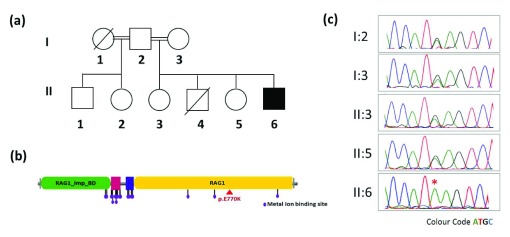
The baby was normal in the perinatal and postnatal period. His birth weight was normal (3.04 kg) and was asymptomatic until 2 months of age. There was a history of admission to pediatric intensive care unit (PICU) and artificial ventilation for severe pneumonia at the age of 2 months. He was admitted for 22 days during that episode. The child had gross motor developmental delay and no adverse events following immunization. He had a male sibling who expired at seven months of age due to persistent pneumonia and two unaffected female siblings, apart from a half-brother and half-sister both of whom were asymptomatic ( Figure 1A).

Figure 1.

Figure 1.

( a) Pedigree of the family ( b) domain mapping of the RAG1 p.E770K on RAG1 protein showing the variation lies on RAG1 domain highlighted with red triangle and ( c) capillary sequencing of the locus in the proband and family members. The homozygous variation c.2308G>A in the proband is marked with an asterisk.

On investigation, the child was found to have hypochromic microcytic anemia, lymphocytopenia, and a normal eosinophil count and platelet count. Liver and renal function tests were normal. The ionized calcium was 0.28mmol/L. Blood culture was positive for Enterobacter species. The child’s mother was HIV ELISA negative and the child had a negative Mantoux test and negative gastric acid AFB stain. His chest X-Ray’s lateral view showed absence of the thymus shadow apart from evidence of bronchopneumonia.

A close workup of the immunoglobulin profile revealed hypogammaglobulinemia-IgA 23 mg%, IgG 44 mg% and IgM 26 mg%. IgE was 1 IU/L. The absolute CD3 count was 464 cells/ul (normal range 1,460–5,440 cells/ul), absolute CD19 lymphocyte count was 12 cells/ul (normal 430–3,300 cells/ul) and absolute NK cell count was 1,328 cells/ul (normal 80–340 cells/ul). Flow cytometry suggested absent B and markedly reduced T cell populations suggestive of B- T- NK+ SCID.

The child was treated with piperacillin (80mg/kg/dose Q8H), vancomycin (15 mg/kg/dose Q6H), dopamine (10 mic/kg/min), intravenous immunoglobulin (IVIG) and other supportive measures and was put on cotrimoxazole (6 mg/kg/day OD) prophylaxis. He was treated with ganciclovir for CMV infection and for staphylococcal pneumonia.

The child was referred for a bone marrow transplant, since SCID is not compatible with life beyond infancy. The patient underwent a matched sibling donor bone marrow transplant at the age of 1 year and 3 months. The conditioning regimen used was Fludarbine 40mg/M2 for 4 days, and Treosulphan 12 gm/M2 for 3 days. GVHD prophylaxis was provided with Methotrexate 10 mg/M2 on days 1, 3 and 6 following transplant, along with Tacrolimus on day 0. Leuconostoc sepsis was treated with intravenous Amoxicillin and Clavulanic acid. Chimerism was assessed using whole blood by fluorescence in situ hybridization (FISH) as the transplant was sex mismatched. At 6 months post- transplant, chimerism was down to 28%, and hence 2 donor lymphocyte infusions were given. The last assessment was done at 1 year by T and B cell markers and serum immunoglobulins, and these were found to be within the normal range. The chilld is now one year three months post-transplant and off all medications including immunosuppressive therapy.

The clinical diagnosis of SCID and family history of sibling death prompted us to investigate the molecular genetic correlates of the disease. Since over 13 genes are implicated in SCID and regular molecular testing was not readily available for the genes, we resorted to whole exome sequencing.

Methods

After obtaining informed consent from the parents, blood was drawn by venipuncture under aseptic precautions. DNA was isolated from whole blood using salting out method 7. Exome capture was performed on DNA using the Illumina Nextera rapid capture expanded exome kit using standard protocols (Illumina Inc USA). We generated 47.95 million paired end reads and an average on target coverage of over 25x on Illumina HiSeq 2500 (Illumina Inc. USA). Alignment was performed using BWA (v0.7.12-r1039) 8 and Stampy (v1.0.20) 9 and variants were called using Platypus (v0.8.1) 10. For the prioritisation of variants, we filtered all homozygous variants, further filtered by an allele frequency of <1% in the 1000 Genome and ExAC. Variants in the 13 genes were prioritised and annotated for their deleteriousness using SIFT, Polyphen and Mutation Taster annotations obtained from annovar 11.

Results

Whole exome sequencing analysis revealed a homozygous missense variation (c.2308G>A) in exon number 2 of recombination activating gene 1 ( RAG1). The variant was predicted to be highly deleterious by SIFT (score 0.000), PolyPhen2 (0.991) and Mutation Taster (1.00). The variation causes an amino acid change p.E770K, which lies on RAG1 domain of the protein ( Figure 1b). The present variation was not found in the 1000 Genome ( http://browser.1000genomes.org/index.html), ExAC ( http://exac.broadinstitute.org/) or internal control database of over 150 exomes from South East Asian ancestry. Incidentally the mutation was previously reported and analysis suggested a significantly reduced recombination activity 12. The variation was further confirmed using targeted PCR amplification around the locus and confirmed by capillary sequencing. The variant was found to be heterozygous in both the parents as well as the surviving siblings ( Figure 1c). The status of the variation could not be ascertained in the sibling who died because no sample was archived and primary immune deficiency was not suspected at the time.

Discussion

Mutations in recombinase activating gene ( RAG1) cause various degrees of severe combined immunodeficiency syndrome (SCID). RAG1 is involved in the V(D)J recombination 1, 2, 13. The child was suspected to have a primary immune deficiency disorder since he had unusually frequent and severe infections and in addition had lost a male sibling due to similar illness. Further, he was born to third degree consanguineous parents. The early onset of symptoms by 2 months of life with increased susceptibility to both bacterial and fungal infections was a pointer to a T cell defect or a phagocytic defect rather than to an antibody deficiency like X linked agammaglobulinemia, which usually presents by 5 to 6 months of age, when maternal antibodies are on the wane 14. The immunoglobulin profile showed that there was also a B cell defect. The low absolute lymphocyte counts coupled with radiological evidence of an absent thymus shadow was proof of a T cell defect as well. Thus, a provisional diagnosis of a severe combined immunodeficiency was made even before the flow cytometry results became available and helped confirm the diagnosis.

The possibility of Omenn syndrome was not considered since there was no history of a rash and there was no lymphadenopathy or hepatosplenomegaly, nor was there eosinophilia in the peripheral smear. X-linked recessive severe combined Immunodeficiency (SCID) is characterized by an elevated percentage of B cells and the absence of B cells in the child ruled this out. Janus kinase 3 (Jak3) deficiency was also not thought of for the same reason. Adenosine deaminase (ADA) deficient SCID is characterized by bony abnormalities including rib cage defects, which were absent.

RAG1 or RAG2 deficiencies are associated with a lack of both B cells and T cells and NK cells are predominant in the circulation 13, 15. With this possibility in mind, and with a view to offer genetic counselling to the family, whole exome sequencing was considered. Whole exome sequencing identified a mutation c.2308G>A p.E770K in RAG1, which was previously reported and shown to significantly reduce recombination activity 12. We feel that whole exome sequencing can have more extensive application in the management of primary immune deficiency in developing countries like India, and can add to rapidly expanding scientific knowledge in this arena.

Ethics approval

The whole exome sequencing was approved by the Institutional Ethical Committee of CSIR - Institute of Genomics and Integrative Biology (IHECC proposal number 8).

Consent

Written informed consent for publication of the patients’ details and/or their images was obtained from the patients/parents of the patient.

Data availability

The data referenced by this article are under copyright with the following copyright statement: Copyright: © 2017 Govindaraj GM et al.

All the raw sequencing data are available at the NCBI Sequence Read Archive ( http://www.ncbi.nlm.nih.gov/sra), accession number SRR4088561.

Acknowledgment

Authors acknowledge support from the GUaRDIAN consortium.

Funding Statement

SS and VS acknowledge funding from the Council of Scientific and Industrial Research (CSIR) India through Grant BSC0212.

The funders had no role in study design, data collection and analysis, decision to publish, or preparation of the manuscript.

[version 2; referees: 2 approved

References

  • 1. Fischer A: Severe combined immunodeficiencies (SCID). Clin Exp Immunol. 2000;122(2):143–9. 10.1046/j.1365-2249.2000.01359.x [DOI] [PMC free article] [PubMed] [Google Scholar]
  • 2. Rivera-Munoz P, Malivert L, Derdouch S, et al. : DNA repair and the immune system: From V(D)J recombination to aging lymphocyte. Eur J Immunol. 2007;37(S1):S71–82. 10.1002/eji.200737396 [DOI] [PubMed] [Google Scholar]
  • 3. Corneo B, Moshous D, Güngör T, et al. : Identical mutations in RAG1 or RAG2 genes leading to defective V(D)J recombinase activity can cause either T-B-severe combined immune deficiency or Omenn syndrome. Blood. 2001;97(9):2772–6. 10.1182/blood.V97.9.2772 [DOI] [PubMed] [Google Scholar]
  • 4. Fischer A, Notarangelo LD, Neven B, et al. : Severe combined immunodeficiencies and related disorders. Nat Rev Dis Primers. 2015;1:15061. 10.1038/nrdp.2015.61 [DOI] [PubMed] [Google Scholar]
  • 5. Zhang W, Cui H, Wong LJ: Application of next generation sequencing to molecular diagnosis of inherited diseases. Top Curr Chem. 2014;336:19–45. 10.1007/128_2012_325 [DOI] [PubMed] [Google Scholar]
  • 6. Sawyer SL, Hartley T, Dyment DA, et al. : Utility of whole-exome sequencing for those near the end of the diagnostic odyssey: time to address gaps in care. Clin Genet. 2016;89(3):275–84. 10.1111/cge.12654 [DOI] [PMC free article] [PubMed] [Google Scholar]
  • 7. Miller SA, Dykes DD, Polesky HF: A simple salting out procedure for extracting DNA from human nucleated cells. Nucleic Acids Res. 1988;16(3):1215. 10.1093/nar/16.3.1215 [DOI] [PMC free article] [PubMed] [Google Scholar]
  • 8. Li H, Durbin R: Fast and accurate short read alignment with Burrows-Wheeler transform. Bioinformatics. 2009;25(14):1754–60. 10.1093/bioinformatics/btp324 [DOI] [PMC free article] [PubMed] [Google Scholar]
  • 9. Lunter G, Goodson M: Stampy: a statistical algorithm for sensitive and fast mapping of Illumina sequence reads. Genome Res. 2011;21(6):936–9. 10.1101/gr.111120.110 [DOI] [PMC free article] [PubMed] [Google Scholar]
  • 10. Rimmer A, Phan H, Mathieson I, et al. : Integrating mapping-, assembly- and haplotype-based approaches for calling variants in clinical sequencing applications. Nat Genet. 2014;46(8):912–8. 10.1038/ng.3036 [DOI] [PMC free article] [PubMed] [Google Scholar]
  • 11. Wang K, Li M, Hakonarson H: ANNOVAR: functional annotation of genetic variants from high-throughput sequencing data. Nucleic Acids Res. 2010;38(16):e164. 10.1093/nar/gkq603 [DOI] [PMC free article] [PubMed] [Google Scholar]
  • 12. Asai E, Wada T, Sakakibara Y, et al. : Analysis of mutations and recombination activity in RAG-deficient patients. Clin Immunol. 2011;138(2):172–7. 10.1016/j.clim.2010.11.005 [DOI] [PubMed] [Google Scholar]
  • 13. Buckley RH: Molecular defects in human severe combined immunodeficiency and approaches to immune reconstitution. Annu Rev Immunol. 2004;22:625–55. 10.1146/annurev.immunol.22.012703.104614 [DOI] [PubMed] [Google Scholar]
  • 14. Shearer WT, Dunn E, Notarangelo LD, et al. : Establishing diagnostic criteria for severe combined immunodeficiency disease (SCID), leaky SCID, and Omenn syndrome: the Primary Immune Deficiency Treatment Consortium experience. J Allergy Clin Immunol. 2014;133(4):1092–8. 10.1016/j.jaci.2013.09.044 [DOI] [PMC free article] [PubMed] [Google Scholar]
  • 15. Notarangelo LD, Kim MS, Walter JE, et al. : Human RAG mutations: biochemistry and clinical implications. Nat Rev Immunol. 2016;16(4):234–246. 10.1038/nri.2016.28 [DOI] [PMC free article] [PubMed] [Google Scholar]
F1000Res. 2017 Oct 10. doi: 10.5256/f1000research.13267.r26544

Referee response for version 2

Paola Itliani 1

No further comments.

I have read this submission. I believe that I have an appropriate level of expertise to confirm that it is of an acceptable scientific standard.

F1000Res. 2017 Oct 3. doi: 10.5256/f1000research.13267.r26546

Referee response for version 2

David K Buchbinder 1

Thank you for the edits to the manuscript. I do think this is a very nice case description which highlights the value of next generation approaches in the diagnosis of primary immunodeficiency disorders.  

My only comments are very minor.  

Introduction:

In the Introduction section I would be review the language and grammar. For example, in the first sentence - "....  of the humoral and/or cellular immune defense mechanism"... sounds awkward. The second sentence may also sound better if you get rid of "the" which starts the sentence. The third sentence where you talk about deficiency of RAG genes being associated with SCID seems awkward. The genes encode the proteins that are deficient. In this case RAG deficiency is associated with T-B-NK+ SCID. I know what you meant, but the sentence is confusing. The next sentence mentions that mutation in RAG1 is associated absence of VDJ recombination. That is not true for all RAG1 variants as you know. Please be careful in your choices of words. The sentence where you talk about an accurate molecular diagnosis being helpful for "genetic counseling for disease" is awkward. Disease what? The last word "gene" (before the sentence "The advent...." should be plural. Lastly, you only need to define the abbreviation RAG1 once.

Case:

You are missing a "he" in the sentence in which you give his birth weight. Define abbreviations like HIV ELISA, AFB, etc. Also, were you able to look at mitogens? That would be useful in defining SCID. What is leuconostoc? What were the doses and timing of the DLI? Any complications such as GVHD? What was the most recent chimerism assessment?  

Methods:

Fine.

Results:

You already defined RAG1.

Discussion:

You already defned RAG1 and SCID. Please use the definition for SCID in the discussion. I would consider "suggested" instead of "was a pointer to". I would get rid of the words "are on the" in front of the word "wane". In the second paragraph you capitalized immunodeficiency. Why? You talk about other forms of SCID such as X-SCID, Jak3, etc. Did you consider other forms of T-B-NK+ SCID? I would consider added this into the discussion.

I have read this submission. I believe that I have an appropriate level of expertise to confirm that it is of an acceptable scientific standard.

F1000Res. 2017 Feb 20. doi: 10.5256/f1000research.10202.r20355

Referee response for version 1

Lennart Hammarström 1

Govindaraj et al. report a typical SCID patient with a known mutation in Rag1 (p.E770K) with a previously known defective recombination activity and thus, there is very little novel information in this manuscript. In fact, the manuscript is already in itself published in F1000 (October 18th, 2016) and available on the web. The mutation should simply be added to existing databases.

Additional comments:

  • The figure should be improved – providing the different domains.

  • The pedigree (inheritance pattern is incomplete) - or simply state that the missing individuals were not sampled.

  • Introduction should be more focussed on Rag1.

  • Immunization schedule (if any) for live vaccines could be of interest for the readers (but not necessary).

  • In the methods section, uniformity should apply (cc, mm ul etc). The number of NK cells appear to be too high (higher than the total lymphocyte count).

  • Gene names should be in italics and transcript IDs should be given.

  • Abbreviations should be correctly used and introduced at the first time of presentation.

I have read this submission. I believe that I have an appropriate level of expertise to state that I do not consider it to be of an acceptable scientific standard, for reasons outlined above.

F1000Res. 2017 Jan 31. doi: 10.5256/f1000research.10202.r19588

Referee response for version 1

David K Buchbinder 1

The authors provide a case report of a patient with severe combined immunodeficiency secondary to RAG deficiency. Whole exome sequencing was utilized to ensure a rapid and accurate diagnosis. Overall, the report is well written. Despite this there are some minor changes that should be considered. I also do not appreciate the novelty of this report and what it adds to the existing literature. The clinical diagnosis of severe combined immunodeficiency is clear. The routine use of next generation sequencing approaches are widely used to provide accurate and rapid diagnoses. 

Introduction

  • Page 3:  I would consider focusing the introduction on the topic of RAG deficiency.

  • Page 3:  I would also note that an accurate and timely diagnosis is vital to the provision of life-saving therapy.

  • Page 3:  At the end of the first paragraph the word “sometime” is missing an “s”.

  • Page 3:  In the last paragraph of the introduction you talk about a genetic variation E770K. A genetic variation in what gene? Please state RAG1.

Case Report

  • Page 3:  When talking about the patient weight, height, and head circumference I would add in percentiles.

  • Page 3:  Please define the abbreviation “PICU”.

  • Page 3:  I would review the units on measurements. I would consider using SI units. For example, please review the use of “mg%”. I would address this issue throughout the manuscript.

  • Page 3:  How was HIV and tuberculosis excluded?

  • Page 3:  Is chest X-Ray correct?

  • Page 3:  Was there any evidence of eosinophilia?

  • Page 4:  For the immunological evaluation -  were antibody responses assessed? Were RA/RO populations assessed? Were T cell responses to mitogens assessed? Was TCR diversity assessed? Was maternal engraftment assessed?

  • Page 4:  Please define the abbreviation “IVIG”.

  • Page 4:  How was the CMV infection identified? Where was the “infection”? How was staph pneumonia identified?

  • Page 4:  I would consider adding a “Methods” section where you talk about the whole exome techniques used and confirmatory sequencing. I don’t really like how it is right in the middle of the case report.

  • Page 4:  In the 4 th paragraph I would italicize “RAG1”. This should be addressed throughout the manuscript.

  • Page 4:  You mention that the mutation was previously reported. This detracts a bit from the novelty. I would also consider noting the RAG activity that was assessed in-vitro from the citation.

Discussion

  • Page 4: You fail to use the abbreviation “SCID” in the first paragraph of the discussion which you defined earlier.

  • Page 4: Please define the abbreviations “XR, Jak3, and ADA”.

  • Page 4: I would include the portion about the transplant course in the case report section and not at the end of the discussion. I would add in additional details as well -  what type of conditioning was used? Graft-versus-host-disease prophylaxis? What was the etiology of the hypertension? PRES? What was the organism isolated during sepsis? Any other post-transplant issues such as GVHD, VOD, etc.? Was engraftment assessed (i.e. lineage specific chimerism)? 

I have read this submission. I believe that I have an appropriate level of expertise to confirm that it is of an acceptable scientific standard, however I have significant reservations, as outlined above.

F1000Res. 2017 Jul 26.
Vinod Scaria 1

The authors provide a case report of a patient with severe combined immunodeficiency secondary to RAG deficiency. Whole exome sequencing was utilized to ensure a rapid and accurate diagnosis. Overall, the report is well written. Despite this there are some minor changes that should be considered. I also do not appreciate the novelty of this report and what it adds to the existing literature. The clinical diagnosis of severe combined immunodeficiency is clear. The routine use of next generation sequencing approaches are widely used to provide accurate and rapid diagnoses.

Introduction

  • Page 3: I would consider focusing the introduction on the topic of RAG deficiency.

Clarification: Suggestions have been incorporated. Introduction is added with focus on RAG1 deficiency.

  • Page 3: I would also note that an accurate and timely diagnosis is vital to the provision of life-saving therapy.

Clarification: Suggestions have been incorporated.

  • Page 3: At the end of the first paragraph the word “sometime” is missing an “s”.

Clarification: Corrections has been incorporated.

  • Page 3: In the last paragraph of the introduction you talk about a genetic variation E770K. A genetic variation in what gene? Please state RAG1.

Clarification: Suggestions have been incorporated.

Case Report

  • Page 3: When talking about the patient weight, height, and head circumference I would add in percentiles.

Clarification: His weight, length and head circumference were below the 3 rd centile as per WHO Child Growth Standards. This is updated in the manuscript.

  • Page 3: Please define the abbreviation “PICU”.

Clarification: Suggestions have been incorporated.

  • Page 3: I would review the units on measurements. I would consider using SI units. For example, please review the use of “mg%”. I would address this issue throughout the manuscript.

Clarification: Ionized calcium was 0.28 mmol/L.

  • Page 3: How was HIV and tuberculosis excluded?

Clarification: The child’s mother was HIV ELISA negative and the child had a negative Mantoux test and negative gastric acid AFB stain.

  • Page 3: Is chest X-Ray correct?

Clarification: Lateral view of the Chest X Ray.

  • Page 3: Was there any evidence of eosinophilia?

Clarification: There was no evidence of eosinophilia

  • Page 4: For the immunological evaluation - were antibody responses assessed? Were RA/RO populations assessed? Were T cell responses to mitogens assessed? Was TCR diversity assessed? Was maternal engraftment assessed?

Clarification: Antibody responses, RA/RO populations, responses to mitogens, TCR diversity and maternal engraftment were not assessed.

  • Page 4: Please define the abbreviation “IVIG”.

Clarification: Suggestions have been incorporated.

  • Page 4:  How was the CMV infection identified? Where was the “infection”? How was staph pneumonia identified?

Clarification: CMV infection was identified by DNA PCR. The child had disseminated CMV infection. Staph. pneumonia was identified by characteristic radiological findings and positive blood culture.

  • Page 4: I would consider adding a “Methods” section where you talk about the whole exome techniques used and confirmatory sequencing. I don’t really like how it is right in the middle of the case report.

Clarification: Suggestions have been incorporated.

  • Page 4: In the 4 th paragraph I would italicize “RAG1”. This should be addressed throughout the manuscript.

Clarification: Suggestions have been incorporated.

  • Page 4: You mention that the mutation was previously reported. This detracts a bit from the novelty. I would also consider noting the RAG activity that was assessed in-vitro from the citation.

Clarification: We are not claiming that the mutation is novel. We are pointing that the variation is so rare that it is reported only once. In-vitro assay for RAG activity from citation is noted down in manuscript.

Discussion

  • Page 4: You fail to use the abbreviation “SCID” in the first paragraph of the discussion which you defined earlier.

Clarification: Suggestions have been incorporated.

  • Page 4: Please define the abbreviations “XR, Jak3, and ADA”.

Clarification: Suggestions have been incorporated.

  • Page 4: I would include the portion about the transplant course in the case report section and not at the end of the discussion. I would add in additional details as well - what type of conditioning was used? Graft-versus-host-disease prophylaxis? What was the etiology of the hypertension? PRES? What was the organism isolated during sepsis? Any other post-transplant issues such as GVHD, VOD, etc.? Was engraftment assessed (i.e. lineage specific chimerism).

Clarification: Suggestion have been incorporated. The hypertension is an inadvertent typographical error. It should read ‘hypotension’. There was no issue of GVHD or VOD.

F1000Res. 2016 Nov 16. doi: 10.5256/f1000research.10202.r17720

Referee response for version 1

Paola Itliani 1

The manuscript “Whole exome sequencing identifies variation c.2308G>A p.E770K in RAG1 associated with B- T- NK+ severe combined immunodeficiency” is the description of a case report, that is a child as a supposed case of SCID. Exactly, the authors found a homozygous variation c.2308G>A by whole exome sequencing and they hypothesized that this variant is associated with the disease. All the sections of the manuscript are well presented and written (title, abstract, content and discussion).

However, there are two points that I would like to comment:

  1. In the introduction the authors wrote that there are over a dozen genes known to be implicated in the disease. So, what about these genes in this case report? By performing a whole exome sequencing, did they also observed the variants of these genes? if yes or not, please comment!

  2. In order to explain the functional consequences of the mutated protein, the authors write “The variation causes an amino acid change p.E770K, which lies on RAG1 domain of the protein……Incidentally the mutation was previously reported and analysis suggested a significantly reduced recombination activity” referring to work of Asai E et al. Clin Immunol. 2011. In Asai’s work the nucleotide mutation is referred as 2420 G>A with a E770K effect. The position of nucleotide between the two works is different, but the authors of the manuscript declare the same position for amino acid change and the same reduced recombination activity. Please, specify/insert the RS ID number of the homozygous variation.

I have read this submission. I believe that I have an appropriate level of expertise to confirm that it is of an acceptable scientific standard, however I have significant reservations, as outlined above.

F1000Res. 2017 Jul 26.
Vinod Scaria 1

The manuscript “Whole exome sequencing identifies variation c.2308G>A p.E770K in RAG1 associated with B- T- NK+ severe combined immunodeficiency” is the description of a case report, that is a child as a supposed case of SCID. Exactly, the authors found a homozygous variation c.2308G>A by whole exome sequencing and they hypothesized that this variant is associated with the disease. All the sections of the manuscript are well presented and written (title, abstract, content and discussion).

However, there are two points that I would like to comment:

1. In the introduction the authors wrote that there are over a dozen genes known to be implicated in the disease. So, what about these genes in this case report? By performing a whole exome sequencing, did they also observed the variants of these genes? if yes or not, please comment

Clarification 1: We have observed the variations in the genes, which are previously reported with primary immunodeficiency. The prioritized variations are shown in table uploaded in answer to reviewer’s comments. (Sent to editor while uploading revised version). Among the variations, the RAG1:c.2308G>A:p.E770K variation was prioritised due to pathogenic and rarity of variation.


2. In order to explain the functional consequences of the mutated protein, the authors write “The variation causes an amino acid change p.E770K, which lies on RAG1 domain of the protein……Incidentally the mutation was previously reported and analysis suggested a significantly reduced recombination activity” referring to work of Asai E et al. Clin Immunol. 2011. In Asai’s work the nucleotide mutation is referred as 2420 G>A with a E770K effect. The position of nucleotide between the two works is different, but the authors of the manuscript declare the same position for amino acid change and the same reduced recombination activity. Please, specify/insert the RS ID number of the homozygous variation.

Clarification 2: In the original publication by Asai et al. 2011, the nucleotide position is mentioned as c.2420G>A. While the position of amino acid is clear from the publication, we are not sure if nucleotide position denotes the position in cDNA or other. In our case, we mentioned the cDNA change as c.2308G>A and amino acid change as p.E770K. We clearly indicated the change in cDNA and it is also the reported nomenclature from database.

For reference, Cosmic mutation ID: COSM3447065.

The rsID for mutation is rs768260595.

Associated Data

    This section collects any data citations, data availability statements, or supplementary materials included in this article.

    Data Availability Statement

    The data referenced by this article are under copyright with the following copyright statement: Copyright: © 2017 Govindaraj GM et al.

    All the raw sequencing data are available at the NCBI Sequence Read Archive ( http://www.ncbi.nlm.nih.gov/sra), accession number SRR4088561.


    Articles from F1000Research are provided here courtesy of F1000 Research Ltd

    RESOURCES