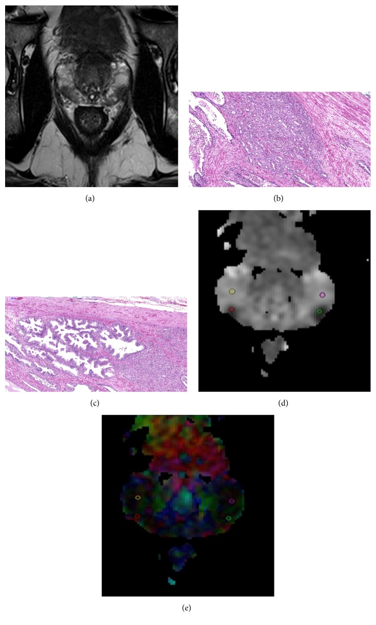
Figure 1.
A 62-year-old patient with two prostate cancer tumor foci in the right and the left peripheral zones both with GS7 (4 + 3). (a) Representative T2-weighted image slice, (b, c) histopathology slices confirming prostate cancer, and (d) MD and (e) FA maps with the ROIs placed for the two tumor foci (solid red line and green line contours) and for the noncancerous tissue, respectively (solid yellow line and purple line contours).

