Figure 5.
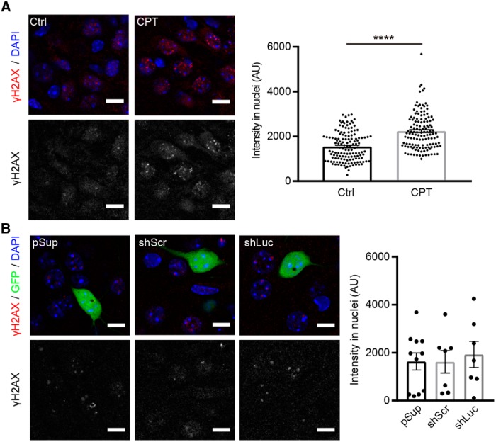
Comparable levels of H2A-X phosphorylation in neurons transfected with different shRNA vectors. γ-H2AX immunoreactivity in shRNA-transfected hippocampal CA1 neurons. A, left, Representative confocal images of immunofluorescence staining against γ-H2AX (red) and DAPI (blue) in CA1 pyramidal neurons. Right, Quantification of γ-H2AX signal intensity in nuclei. Treatment with 10 μM camptothecin (CPT) for 6 h increased the signal of γ-H2AX in nuclei of neurons in organotypic slice cultures, confirming the specificity of the anti-γ-H2AX antibody. AU, arbitrary units. Number of cells/mice: mock control (Ctrl), 140/3; CPT, 133/3. B, left, Representative confocal images of immunofluorescence staining against γ-H2AX (red), GFP epifluorescence (green) and DAPI (blue) in pSup-, shLuc-, and shScr-transfected neurons. Right, Quantification of γ-H2AX signal intensity in nuclei. All transfected neurons exhibited comparable levels of γ-H2AX signal. Number of cells/mice: pSup, 11/3; shScr, 7/4; shLuc, 7/3. Scale bars: 10 μm.

