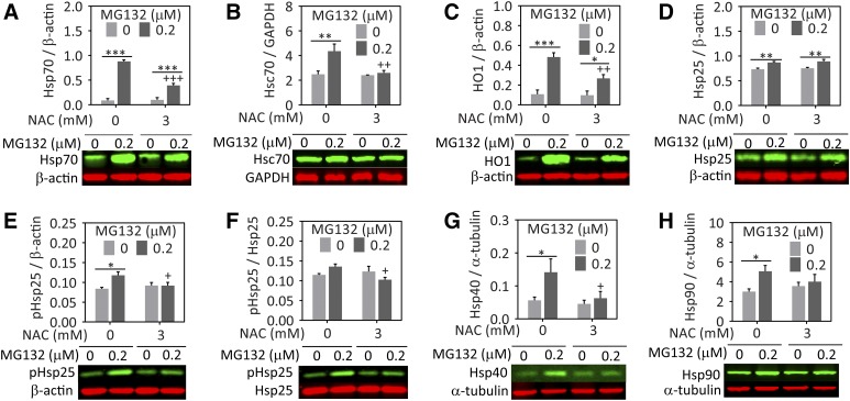
Fig. 4.
NAC attenuates the MG132-induced increase in heat shock proteins in astrocytes. Primary cortical astrocytes were treated with MG132 or vehicle in the presence of NAC or vehicle. Lysates were collected 24 hours later and heat shock proteins were measured by western blot analyses. Representative western blot images are included below each graph. (A) Hsp70, (B) Hsc70, (C) HO1 (or heat shock protein 32), (D) heat shock protein 25 (Hsp25), (E and F) phosphorylated Hsp25, (G) heat shock protein 40 (Hsp40), and (H) heat shock protein 90 (Hsp90). Protein levels are expressed as a function of the loading controls β-actin, glyceraldehyde-3-phosphate dehydrogenase (GAPDH), or α-tubulin, depending on the species of the antibody for the protein of interest and its molecular weight. Infrared images are pseudocolored green and red. Shown are the mean and S.E.M. values of 3–5 independent experiments. *P ≤ 0.05; **P ≤ 0.01; ***P ≤ 0.001 versus 0 µM MG132; +P ≤ 0.05; ++P ≤ 0.01; +++P ≤ 0.01 versus 0 mM NAC, two-way analysis of variance followed by the Bonferroni post hoc test.

