Fig. 3.
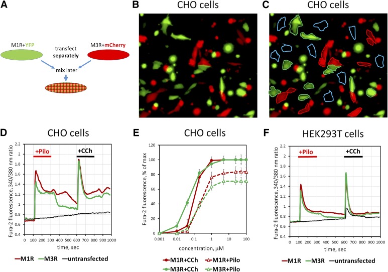
Pilocarpine stimulates free Ca2+ mobilization in cells overexpressing M3R. CHO-K1 or HEK293T cells were transiently transfected with M3R, M1R, and fluorescent proteins. (A) Schematic of the experiment. Cells transfected to express M1R with eYFP or M3R with mCherry (red and green) are mixed and plated on coverslips. They are subsequently loaded with fura-2, AM and analyzed for sensitivity to cholinergic stimulation. (B and C) A representative image (Original magnification, 200×) of the mixed cell population. Red cells are cotransfected with plasmids encoding M3R and mCherry, and green cells express eYFP together with M1R. (C) Illustration of selection of the regions of interest to collect data on Ca2+. Blue traces denote cells that do not express fluorescent proteins and are visualized by furae staining alone. See Materials and Methods for additional details. (D) Free Ca2+ responses to 10 μM pilocarpine and 10 μM CCh. Traces represent the average of responses recorded from 20 to 30 individual cells per region of interest. Green trace corresponds to the data from M1R-expressing cells, red shows M3R, and black shows untransfected cells. Data shown are representative of at least three such experiments done with independent transfections. (E) Amplitude of Ca2+ responses was measured at the indicated concentrations of CCh or pilocarpine. (F) Experiment on HEK293T cells performed essentially as that done on CHO-K1 cells (A–D). Note that there is a response of untransfected cells to CCh but not to pilocarpine. Representative of two independent transfection experiments.

