Fig. 8.
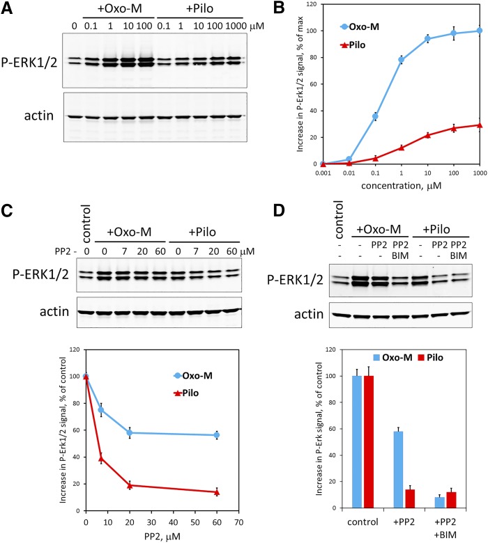
Pilocarpine stimulates ERK1/2 phosphorylation in MIN6 cells via a PP2-sensitive pathway. (A) MIN6 cells were serum-starved for 4 hours and then stimulated for 5 minutes with the indicated concentrations of Oxo-M or pilocarpine (Pilo). The amount of phosphorylated ERK1/2 was determined by Western blot using anti-P-ERK1/2(T202/Y204) antibody. The same membrane was also stained with an anti-actin antibody used for signal normalization. Shown is a representative immunoblot. (B) Quantification of ERK1/2 phosphorylation in response to stimulation with Oxo-M (blue) or pilocarpine (Pilo, red) was done as described in Materials and Methods. Data show mean ± S.D. from three independent experiments. (C) PP2, a Src family kinase blocker, inhibits pilocarpine- and Oxo-M-stimulated ERK1/2 phosphorylation. MIN6 cells were serum-starved and preincubated with the indicated concentrations of PP2 for 4 hours. Then they were stimulated for 5 minutes with either 1 μM Oxo-M (blue) or 100 μM pilocarpine (Pilo, red). ERK1/2 phosphorylation was determined as in (A and B). Shown is a representative immunoblot and data quantification (mean ± S.D. from three independent experiments). (D) A PKC inhibitor bisindolylmaleimide I (BIM, 10 μM) almost completely blocked Oxo-M-stimulated ERK1/2 phosphorylation when combined with PP2 (60 μM). The experiment was performed and quantified as in (C). Data show mean ± S.D. from three independent experiments.

