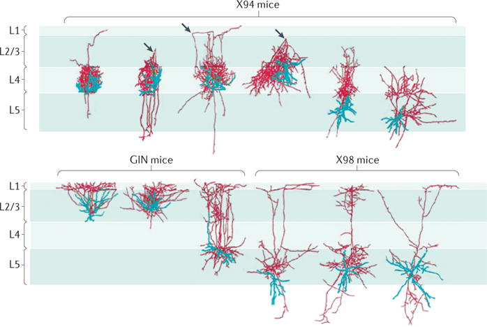
Figure 1.

Three-dimensional morphological reconstructions of SST interneurons in the primary somatosensory cortex of different transgenic mouse lines. Somatostatin-expressing (SST) neuron diversity is highlighted by the differences in anatomical properties of cells within and across layers. Cell bodies and dendrites are shown in blue, and axons are shown in red. The three arrowheads in the top row point to a turning point of the axon, from the upper layers back to layer 4 (L4). The top panel shows neurons in the X94 line. The bottom panel shows neurons in the green fluorescent protein (GFP)-expressing inhibitory neuron (GIN) line and in the X98 line. Note that in the GIN and X98 lines, both L2/3 and L5 SST neurons have an axonal branch that ascends and prominently elaborates in L1, as well as substantial branching within the ‘home’ layer that contains the cell body. L4 SST neurons may also have an axonal branch that ascends to L1, although most of the axon is concentrated in L4. This selective axonal targeting to L1 is similar to that observed in hippocampal stratum oriens–lacunosum moleculare (OLM) neurons, in which the SST cell body lies in the stratum oriens, and the axon elaborates in the lacunosum moleculare. Dense, lamina-specific axons from SST neurons suggest an important role for these cells in regulating synapses that lie within this layer through either GABA type A receptor (GABAAR)- or GABABR-mediated mechanisms. Figure is republished with permission of Society for Neuroscience, from Distinct subtypes of somatostatin-containing neocortical interneurons revealed in transgenic mice. Ma, Y., Hu, H., Berrebi, A. S., Mathers, P. H. & Agmon, A., J. Neurosci. 26 (19) 5069–5082 (2006); permission conveyed through Copyright Clearance Center, Inc.
