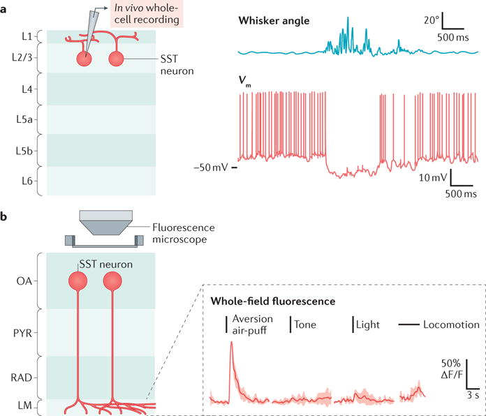
Figure 2.

Regulation of SST-neuron activity during movement and sensation. The activity of somatostatin-expressing (SST) neurons can be up- or downregulated during different activities and behaviours, and in response to different stimuli. a | Schematic of the configuration (left panel) used for in vivo whole-cell recording (right panel; red trace) from a layer 2/3 (L2/3) SST neuron from mouse barrel cortex, during periods of quiet resting or whisking activity (right panel; blue trace). Whisking is associated with hyperpolarization of the SST neuron. b | Schematic of the calcium imaging configuration (left panel) used for the whole-field measurement of calcium transients in different cell types in different layers of the hippocampus — in this case, SST neurons with cells bodies in the stratum oriens–alveus (OA) that project to the stratum lacunosum moleculare (LM), known as OLM cells. Example trials (right panel) of Ca2+ transients in the LM during sensory stimuli and locomotion show that SST neurons increase their activity during presentation of the unconditioned stimulus (an aversive air-puff), but not to other sensory stimuli or during locomotion. PYR, stratum pyramidale; RAD, stratum radiatum; Vm, membrane potential. Part a is adapted from REF. 52, Nature Publishing Group. Part b is from Lovett-Barron, M. et al. Dendritic inhibition in the hippocampus supports fear learning. Science 343, 857–863 (2014); reprinted with permission from AAAS.
