. 2017 Apr 13;8(6):4223–4230. doi: 10.1039/c7sc00641a

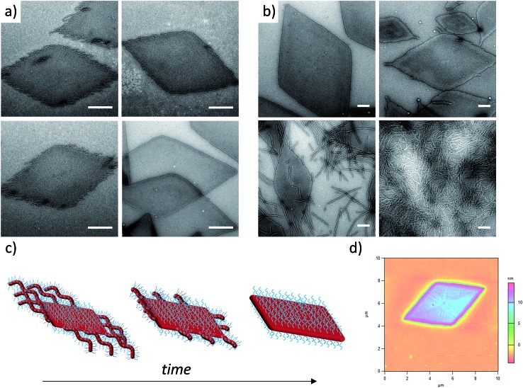
Fig. 2. (a) TEM micrographs of corona–core ratio 20 : 1 PDMA1000-b-PLLA48 self-assembled in ethanol at 90 °C for 2 h (top left), 4 h (top right), 6 h (bottom left) and 8 h (bottom right) before cooling to room temperature. (b) TEM micrographs of a series of PDMAm-b-PLLA48 block copolymers of corona–core ratios of 20 : 1 (m = 1000, top left), 12.5 : 1 (m = 600, top right), 5 : 1 (m = 250, bottom left), and 3 : 1 (m = 150, bottom right). Samples were self-assembled in ethanol at 90 °C for 8 h and cooled to room temperature. All samples were stained with uranyl acetate. Scale bar = 1 μm. (c) Schematic of diamond platelet formation kinetics. (d) AFM of diamond platelet assembled from 20 : 1 corona–core ratio diblock copolymer.

Fig. 2