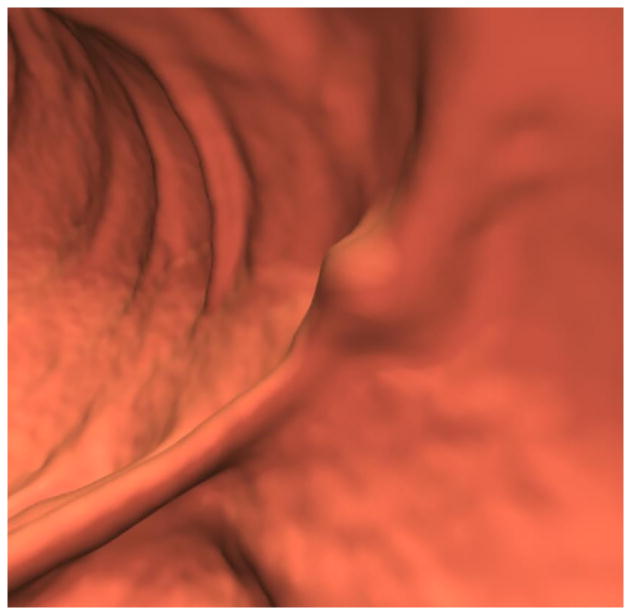
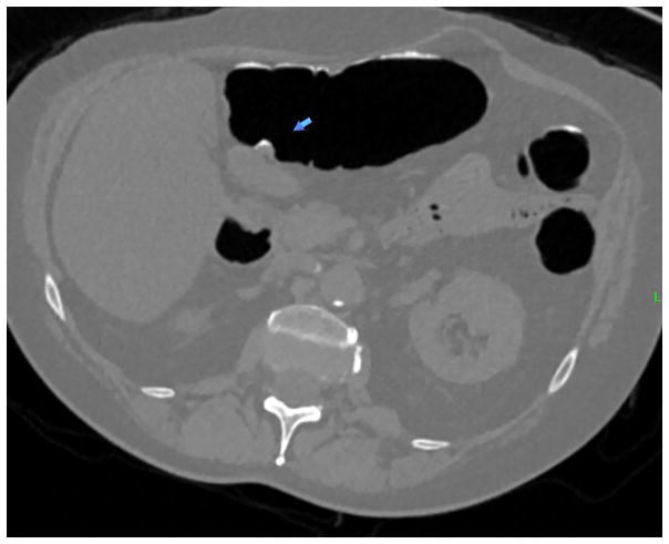
Figure 1. Sub-cm hyperplastic polyp at the colonic anastomosis (1.4 years after right hemicolectomy for CRC) in 70-year-old woman.

A and B, 3D (A) and 2D (B) CTC images show a focal 8-mm soft tissue polyp (arrow) at the anastomosis. The high attenuation seen at the edge of the lesion on 2D represents either oral contrast coating or calcification involving a suture granuloma. Given the OC findings, the latter is favored.
C, Image from same-day OC confirms a polyp at the anastomosis, with adjacent suture material. The lesion proved to be a hyperplastic polyp at surgical pathology.


