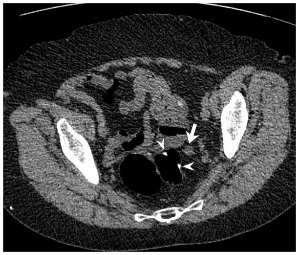
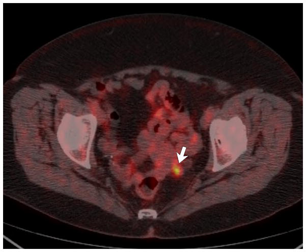
Figure 2. Peri-anastomotic cancer recurrence (12 months after resection of T4aN0M0 sigmoid cancer) in 76-year-old woman with slightly elevated CEA level (7 ng/mL).
A and B, 2D low-dose unenhanced prone (A) and post-contrast supine (B) CTC images show an irregular 1 cm soft tissue nodule (arrows) adjacent to the colonic anastomosis (arrowheads). Note the peripheral enhancement of the nodule on the contrast-enhanced view, as well as collapse of the anastomosis. The anastomosis was deemed normal at OC (not shown).
C, Fused image from PET/CT obtained after the CT/CTC study shows that the lesion is hypermetabolic, which proved to be local recurrence.


