Fig. 3.
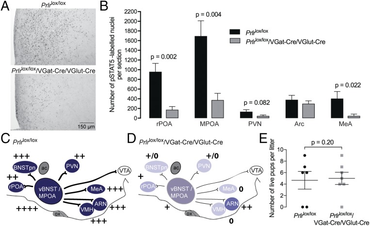
Effect of conditional deletion of Prlr in both GABA and glutamate neurons. (A) Prolactin-induced pSTAT5 (black nuclear staining) in the MPOA showing functional loss of Prlr signaling in combined GABA and glutamate Prlr KO mice (Prlrlox/lox/VGat-Cre/VGlut-Cre) together with quantification of the numbers of pSTAT5-positive cells in selected brain regions (n = 4–5). (B) Note the significant loss of prolactin-responsive cells in most brain regions. This is represented schematically in the diagrams of the brain in the sagittal plane (C and D), with intensity of color representing levels of Prlr expression and + symbols representing numbers of prolactin-induced pSTAT5 neurons detected in each region. Note the marked loss of Prlr expression throughout that neural network. E shows pup survival measured on day 3 of lactation. Although some mothers abandoned their litters in each group, there was no significant effect of Prlr deletion on postpartum maternal behavior compared with Cre-negative controls. ac, anterior commissure; ARN, arcuate nucleus; BNSTpn, bed nucleus of the stria terminalis principle nucleus; LS, lateral septum; MeA, medial amygdala; ox, optic chiasm; PAG, periaqueductal gray; PVN, paraventricular nucleus; rPOA, rostral preoptic area; SON, supraoptic nucleus; vBNST, ventral bed nucleus of the stria terminalis; VTA, ventral tegmental area.

