Fig. S2.
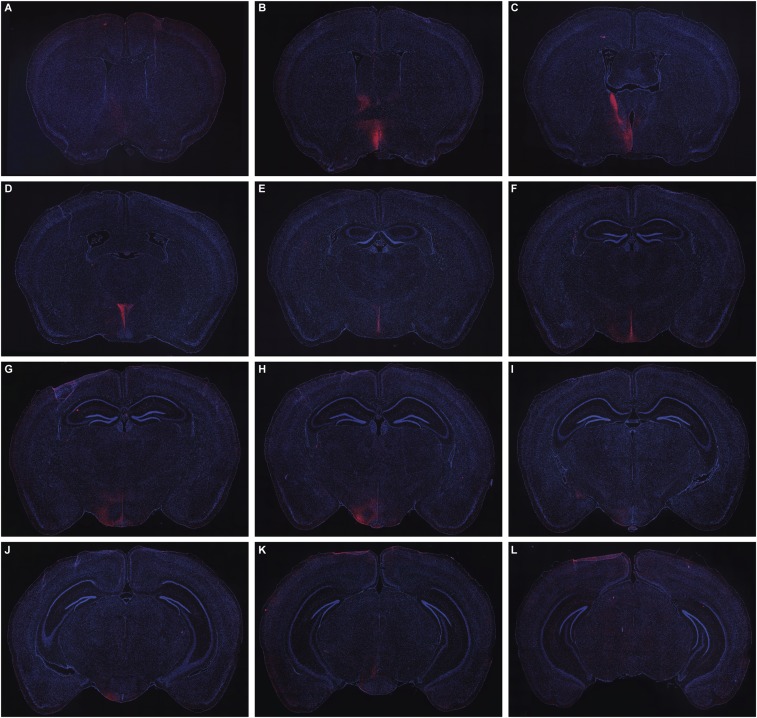
Serial sections through a single animal showing the distribution of mCherry-labeled fibers (red) derived from prolactin-responsive neurons in the MPOA after administration of AAV5-EF1a-DIO-hChR2(H134R)-mCherry-WPRE-pA into the MPOA of Prlr-iCre/eR26-\x{03c4}GFP mice. Note: These are the same sections as Fig. S1, using the same magnification. Images were captured using a 10× objective using a Zeiss Axio Imager2 microscope with a motorized stage, with multiple images combined to form a composite image using the MozaiX module in the Axiovision software. Note the predominantly ipsilateral distribution extending from the rostral preoptic area (A), BNST (B and C), PVN (D), periventricular nucleus (D–F), ARN and VMH (G–I), and VTA (J–L).

