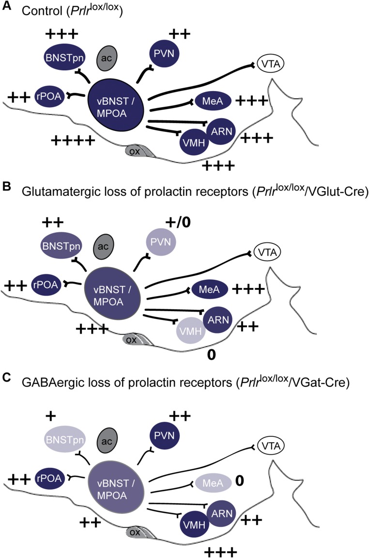
Fig. S3.
Conditional deletion of the Prlr from glutamate or GABA neurons. Schematic representation of levels of Prlr expression in the maternal circuit in the sagittal plane, with intensity of color representing levels of Prlr expression and + symbols representing numbers of prolactin-induced pSTAT5 neurons detected in each region (A). Note the reduced expression of Prlr and prolactin-induced pSTAT5 in specific regions after Cre-mediated deletion of Prlr in glutamate (VGlut-Cre; B) or GABA (VGat-Cre; C). ac, anterior commissure; ARN, arcuate nucleus; BNSTpn, bed nucleus of the stria terminalis principle nucleus; MeA, medial amygdala; MPOA, medial preoptic nucleus; ox, optic chiasm; PVN, paraventricular nucleus; rPOA, rostral preoptic area; vBNST, ventral bed nucleus of the stria terminalis; VTA, ventral tegmental area.

