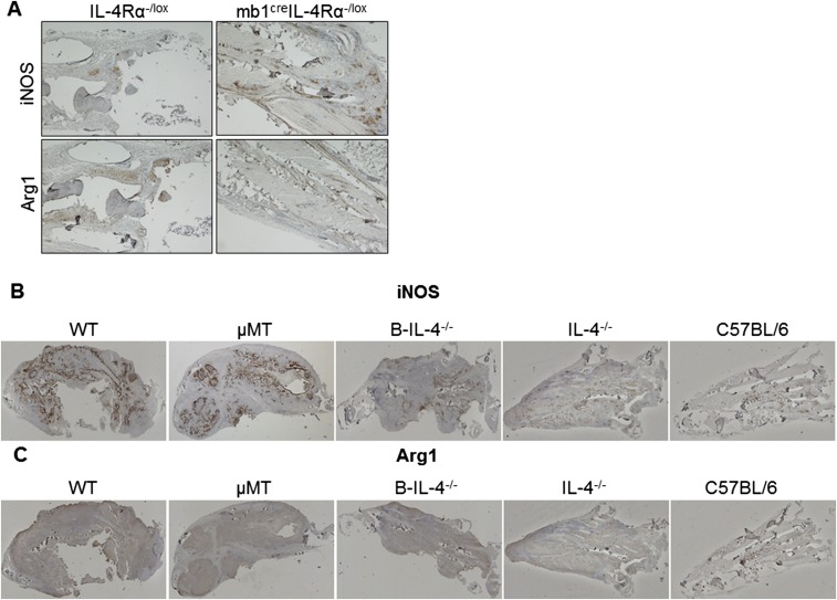
Fig. S3.
iNOS and arginase staining in footpads of mice infected with L. major LV39. (A) iNOS and arginase 1-positive staining in formalin-fixed footpad sections of LV39-infected mb1creIL-4Rα–/lox BALB/c and control mice, and (B and C) respective chimeric mice (Fig. S7) by immunohistochemical staining. Representative images were acquired at 4× objective lens (A) and 2× objective lens (B and C) using a Nikon 90i microscope and scanned for quantification of positive staining expressed as a ratio of iNOS+/Arg-1+ (Figs. 2G and 5I), serving as a proxy for classical and alternative activation, respectively. (Scale bar: A, 500 µm; B and C, 1,000 µm.)

