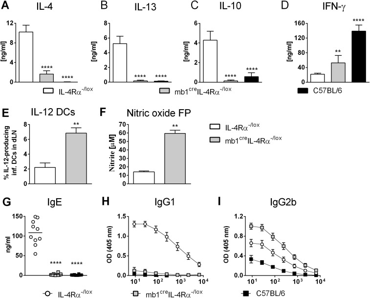
Fig. S4.
Impaired Th2 cytokine responses and killing effector functions in mb1creIL-4Rα–/lox mice infected with L. major IL81. (A–C) mb1creIL-4Rα–/lox BALB/c and control mice were infected s.c. with 2 × 10⁵ L. major IL81 promastigotes into the hind footpad. At week 6 postinfection, total LN CD4+ T cells were restimulated for 72 h with fixed APCs and SLA. The production of IL-4 (A), IL-13 (B), IL-10 (C), and IFN-γ (D) in cell supernatants was determined by ELISA. (E) CD11chiMHCIIhiCD11b+Ly6chi inflammatory DCs in the LN were analyzed for the production of IL-12 by intracellular FACS staining at week 6 after IL81 infection. (F) Production of NO in total footpad cells. Total cells were isolated from footpads at week 6 after IL81 infection and stimulated with 10 ng/mL LPS for 72 h. Production of NO was determined in cell supernatants. (G–I) At week 6 post-IL81 infection, total IgE (G), antigen-specific IgG1 (H), and IgG2b (I) antibody isotypes were quantified from infected sera by ELISA. A representative of two individual experiments is shown with mean values ± SEM. Statistical analysis was performed defining differences to littermate IL-4Rα–/lox BALB/c control mice as significant (**P ≤ 0.01, ****P ≤ 0.0001).

