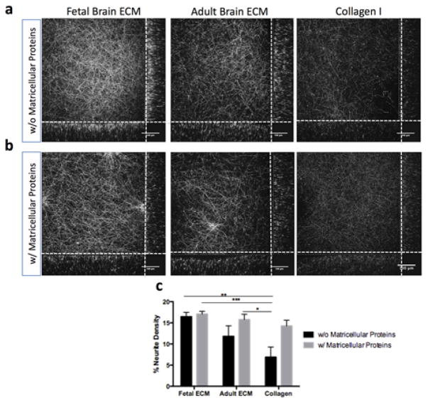
Figure 4.
Primary rat neurons in silk-collagen 3D bioengineered brain model in the presence of ECM and matricellular proteins. (a) Growth of primary cortical rat neurons (embryonic day 18) in 3D silk scaffold infused with collagen I gel or collagen I supplemented with fetal or adult brain ECM at a concentration of 1000μg/ml. Neurites stained by β-III Tubulin at 2wk time point. (b) Growth of primary cortical rat neurons in 3D silk scaffold infused with collagen I gel or collagen I supplemented with fetal or adult brain ECM at 1000μg/ml of gel. The culture media was additionally supplemented with astrocyte-released matricellular proteins (SPARC, Hevin, TSP2). Neurites stained by β-III Tubulin at 2wk time point. (c) Average neurite density per 2D plane of corresponding 3D Z-stack as quantified by using a custom image analysis code; ***p=0.001, **p<0.005, *p<0.007; n=5; collagen I as control

