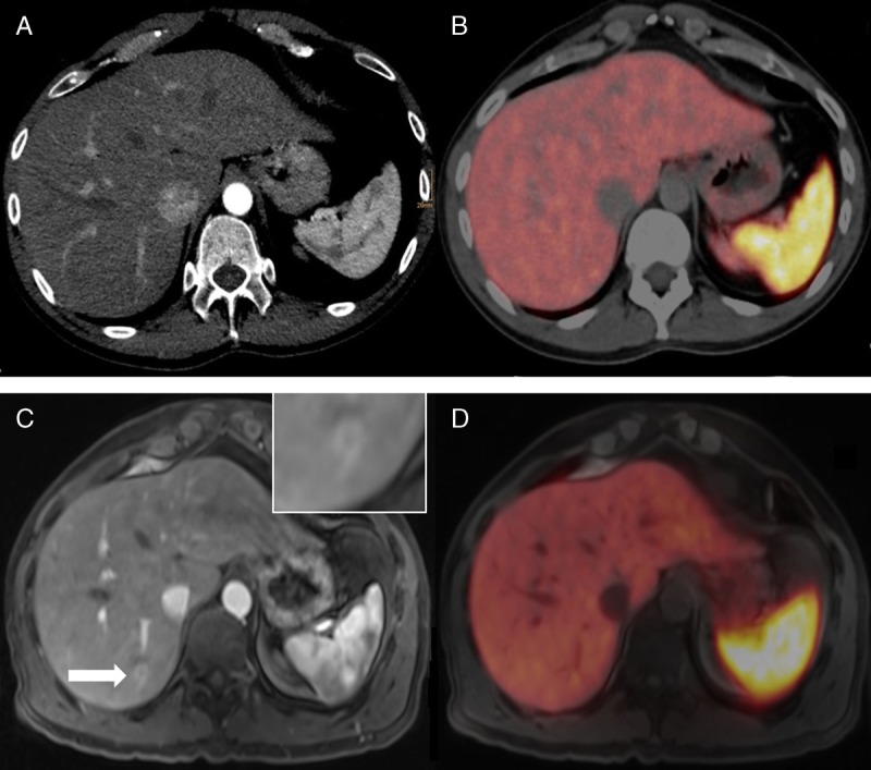
FIGURE 2.

A 37-year-old man after surgically resected primary NET (G1) of the small bowel. In the arterial contrast medium phase in PET/CT (A), no focal liver lesion could be detected. In the corresponding fused PET images (B), no suspicious focal uptake was found in the liver parenchyma. PET/MRI revealed a small lesion in liver segment VI (C, T1 VIBE, arterial contrast medium phase) and focal uptake on the corresponding fused PET images (D).
