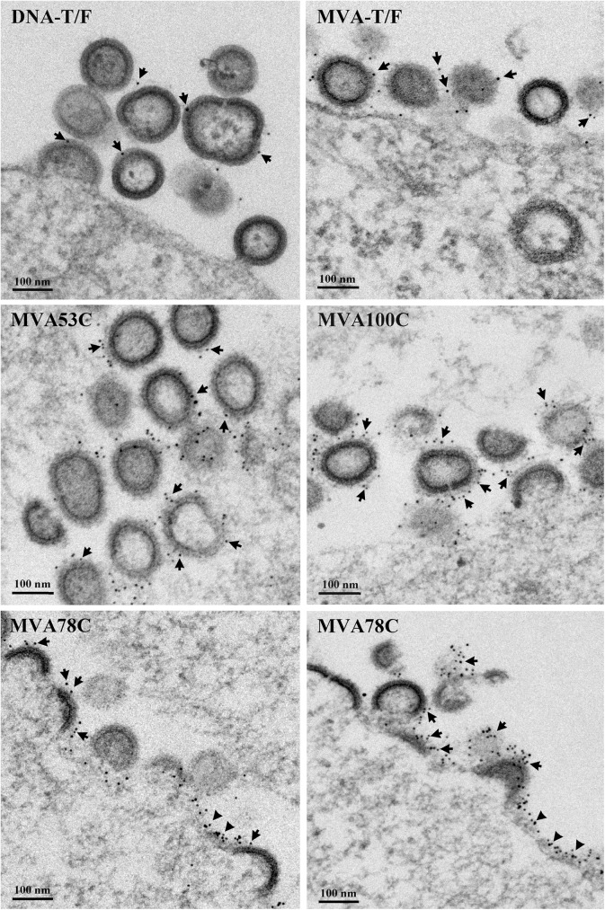
Fig 2. Electron micrographs of VLPs expressed by the DNA and MVA vaccines.
Thin section electron micrographs were immunogold stained for Env using the PGT145 and PGT151 Ab that bind native trimers (see methods). The DNA vaccine is expressed in transiently transfected 293T cells and the MVA vaccine in infected DF1 cells. The VLPs being analyzed and nanometer (nm) size markers are indicated in the panels. Arrows, indicate examples of immunogold staining on VLPs. The triangles point to examples of immunogold staining on the plasma membranes of vaccine infected cells.

