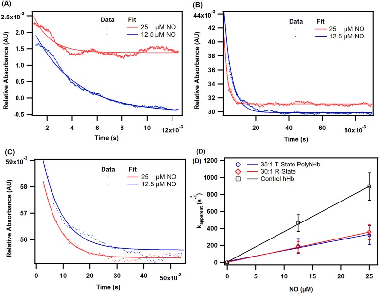
Fig 7. Time courses for the NO deoxygenation reaction.
Time courses for the NO dioxygenation reaction with oxygenated (A) hHb, (B) 30:1 R-state PolyhHb, and (C) 35:1 T-state PolyhHb. Dots represent experimental data and the corresponding solid lines of the same color represent curve fits. Experimental data shows an average of 7–10 kinetic traces. The reactions were monitored at 420 nm and 20°C. PBS (0.1 M, pH 7.4) was used as the reaction buffer. (D) Comparison of NO dioxygenation rates of hHb, 35:1 T-state PolyhHb, and 30:1 R-state PolyhHb. The error bars represent the standard deviation from 15 replicates.

