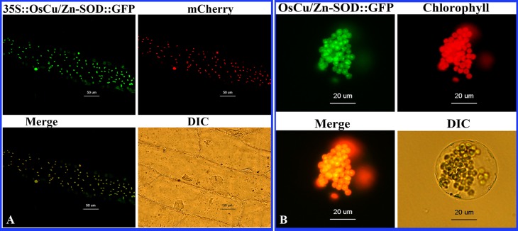
Fig 3. Subcellular localization of the 35S::OsCu/Zn-SOD::GFP fusion protein.
(A) To interpret the color references in this figure legend, the reader should refer to the web version of this article. Fluorescence of OsCu/Zn-SOD-GFP fusion protein in onion; merged image of the subcellular localization of the chloroplast marker mcherry; superposition of green fluorescence and mCherry; DIC, bright field. (B) Targeting of GFP fusion proteins to Arabidopsis protoplast. Fluorescence of OsCu/Zn-SOD-GFP fusion protein in a protoplast derived from an Arabidopsis mesophyll cell; autofluorescence of chlorophyll; merged images from green and red channels; protoplast viewed under white light.

