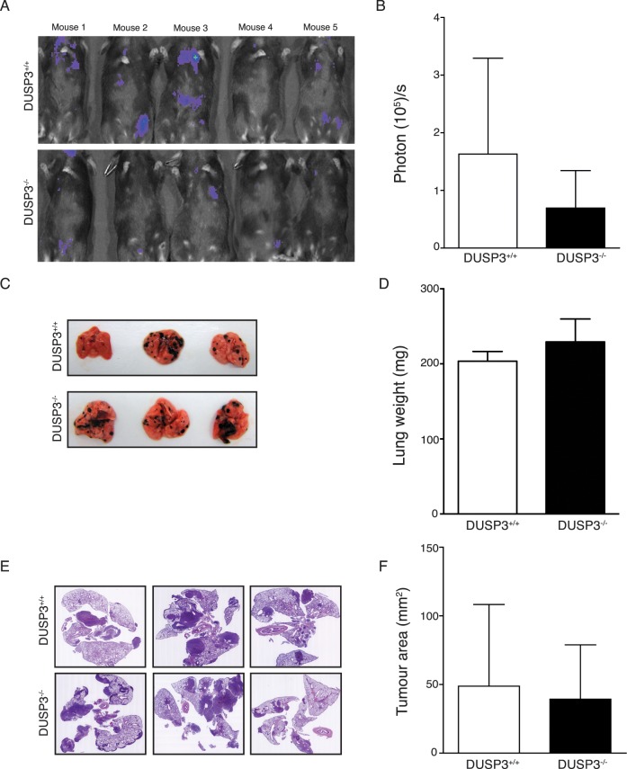
Fig 2. DUSP3 deletion does not impact experimental B16 metastasis growth.
B16 tumour growths were monitored by xenogen bioluminescence imaging. Tumours were established by i.v. injection of 106 B16-Luc+ cells to DUSP3+/+ and DUSP3-/- mice. (A) Representative xenogen imaging results and (B) quantitative xenogen bioluminescence imaging data (day 14). (C) Representative lung macroscopic view and (D) comparison of lung weights from DUSP3+/+ and DUSP3-/- mice. (E) Hematoxylin eosin staining of lung sections from each experimental group. (F) Comparison of tumour areas from each group. Student t-test was used for (B) and (D) and Mann-Whitney test was used for (F). *p < 0,05, **p < 0.01. 5 mice in each group were used for each experiment. Data shown are representative of 4 different experiments.

