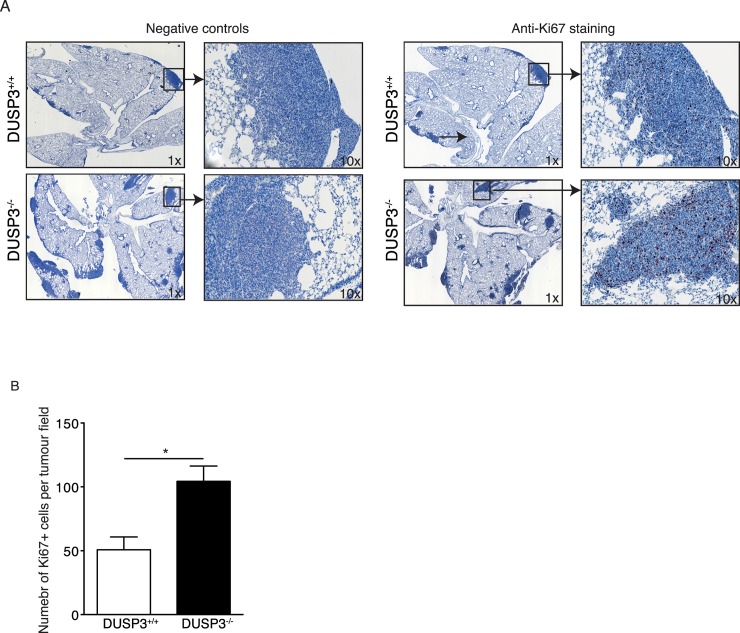
Fig 8. Increased LLC tumour mass in DUSP3-/- mice lungs is associated with increased in situ proliferation of these cells.
Sections of 5-μm thickness were cut from DUSP3-/- and DUSP3+/+ LLC bearing lungs embedded in paraffin blocks. Immunohistochemistry for Ki67 was carried out. Revelation was performed using AEC+ Red. Negative control sections were traited similarly except that they were not stained with anti-KI67. (A) repesentative images from each group of mice are shown at 1x and 10x magnifications. Dark color indicate positive staining for Ki67. (B) Quantification of the positively stained cells from the entire tumor represented in the section using Image J software. Student-t-test was used for statistical analysis. *p < 0.05. Data shown are representative of 5 different sections scanned from 5 individual mice from each genotype.

